Utility of the inversion scout sequence (TI scout) in diagnosing myocardial amyloid infiltration
- PMID: 22527255
- PMCID: PMC3586540
- DOI: 10.1007/s10554-012-0042-4
Utility of the inversion scout sequence (TI scout) in diagnosing myocardial amyloid infiltration
Abstract
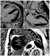
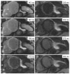
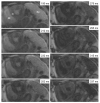
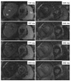
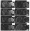
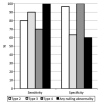
To evaluate the utility of inversion scout (TI-scout) obtained during cardiac magnetic resonance imaging (CMR) in diagnosing myocardial amyloid infiltration. A retrospective analysis of CMR exams in 39 patients (24 males, age range 29-77 years) was performed. Imaging was performed on a 1.5T system, and included steady state cine, post contrast TI-scout and delayed enhancement images. Evaluations included studies in 13 patients with myocardial amyloidosis and 26 patients without myocardial amyloidosis. To characterize abnormal nulling, the time to myocardial nulling on the TI scout was compared to the null times of the blood pool and spleen for each scan. The sensitivity and specificity of different tissue nulling abnormalities for myocardial amyloidosis were computed. The null times of tissues in 18/26 (69%) patients in the non-amyloid group followed a consistent order with the blood pool null time preceding the myocardial nulling which was equal to that of splenic nulling (Type 1 pattern). This order differed in all 13 patients with myocardial amyloidosis described as three non-mutually exclusive nulling categories: 10 patients had myocardial null time preceding or coincident with blood pool (Type 2 pattern); in 11 patients myocardial null time was non-coincident with splenic nulling (Type 3 pattern); and in 8 patients myocardial null time was non-coincident with both blood pool AND splenic nulling (Type 4 pattern). While no patient exhibited Type 4 nulling pattern in the non-amyloid group, 1/26 patient had a Type 2 and 7/26 patients had a Type 3 nulling pattern. A sensitivity of 100% was obtained when either Type 2 OR Type 3 nulling was present while a specificity of 100% was obtained when both Type 2 AND Type 3 nulling were present together (Type 4 pattern). Our study demonstrates that the pattern of nulling on the TI scout sequence CMR has potential diagnostic utility for the presence of myocardial amyloidosis. The temporal pattern of myocardial, blood pool and splenic nulling needs to be carefully evaluated on the TI scout sequence and could prove useful in other infiltrative cardiomyopathies.
Figures

References
-
- Falk RH, Skinner M. The systemic amyloidoses: an overview. Adv Intern Med. 2000;45:107–137. - PubMed
-
- Rahman JE, Helou EF, Gelzer-Bell R, Thompson RE, Kuo C, Rodriguez ER, Hare JM, Baughman KL, Kasper EK. Non-invasive diagnosis of biopsy proven cardiac amyloidosis. J Am Coll Cardiol. 2004;43:410–415. - PubMed
-
- Syed IS, Glockner JF, Feng DL, Araoz PA, Martinez MW, Edwards WD, Gertz MA, Dispenzieri A, Oh JK, Bellavia D, Tajik AJ, Grogan M. Role of cardiac magnetic resonance imaging in the detection of cardiac amyloidosis. J Am Coll Cardiol Cardiovasc Imaging. 2010;3:155–164. - PubMed
-
- Vanden Driesen RI, Slaughter RE, Strugnell WE. MR findings in cardiac amyloidosis. AJR. 2006;186:1682–1685. - PubMed
-
- Maceira AM, Joshi J, Prasad SK, et al. Cardiovascular magnetic resonance in cardiac amyloidosis. Circulation. 2005;111(2):186–193. - PubMed
Publication types
MeSH terms
Grants and funding
LinkOut - more resources
Full Text Sources
Other Literature Sources
Medical

