

Review
doi: 10.1007/s00018-012-0984-7.
Epub 2012 Apr 19.
The intestinal epithelium tuft cells: specification and function
Affiliations
- PMID: 22527717
- PMCID: PMC3417095
- DOI: 10.1007/s00018-012-0984-7
Item in Clipboard
Review
The intestinal epithelium tuft cells: specification and function
Cell Mol Life Sci.
2012 Sep.
Abstract
The intestinal epithelium, composed of at least seven differentiated cell types, represents an extraordinary model to understand the details of multi-lineage differentiation, a question that is highly relevant in developmental biology as well as for clinical applications. This review focuses on intestinal epithelial tuft cells that have been acknowledged as a separate entity for more than 60 years but whose function remains a mystery. We discuss what is currently known about the molecular basis of tuft cell fate and differentiation and why elucidating tuft cell function has been so difficult. Finally, we summarize the current hypotheses on their potential involvement in diseases of the gastro-intestinal tract.
Figures
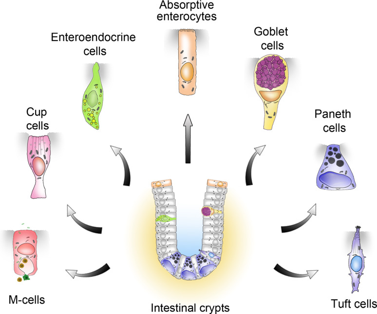
Schematic representation of the known intestinal epithelial cell types generated from Lgr5-expressing crypt base columnar stem cells
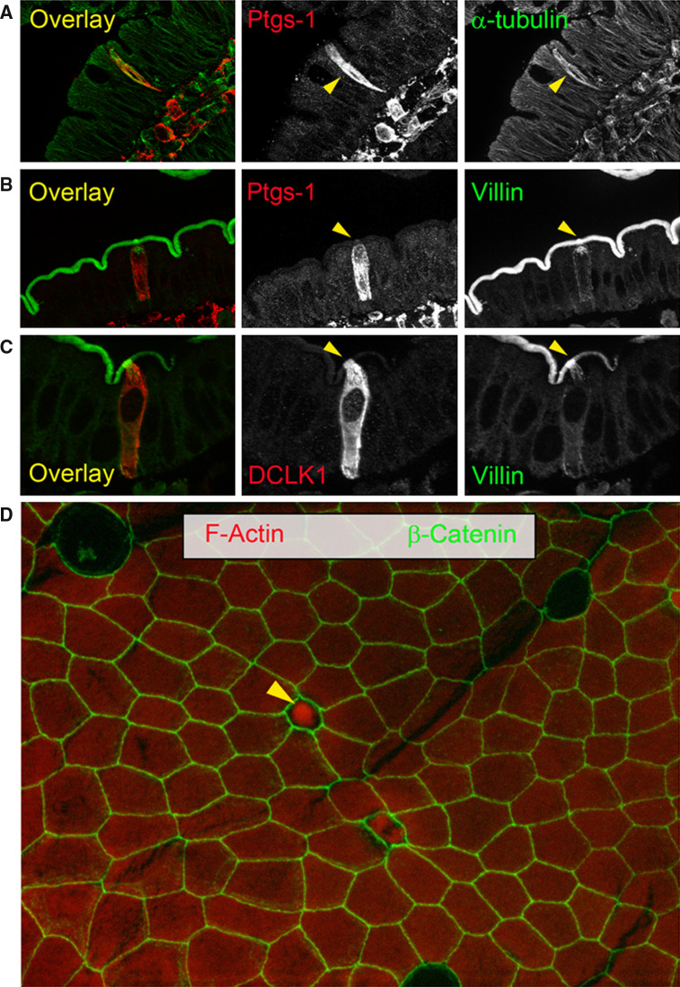
Unique molecular and structural features distinguish tuft cells from the other intestinal epithelial cells. a–d Tuft cells are indicated by arrowheads. a α-tubulin staining (green) highlights the dense microtubule network of a tuft cell co-stained for Ptgs-1 (red). b Tuft cell co-stained for Ptgs-1 (red) and villin (green). c Tuft cell co-stained for DCLK1 (red) and villin (green). Note the increased Villin immunoreactivity of the apical pole and rootlets (yellow
arrowhead). d Surface epithelium of a villus (whole mount) stained for β-catenin (green) and phalloidin (red). The apical part of a tuft cell highly immunoreactive for F-actin sticks out into the lumen (yellow
arrowhead)
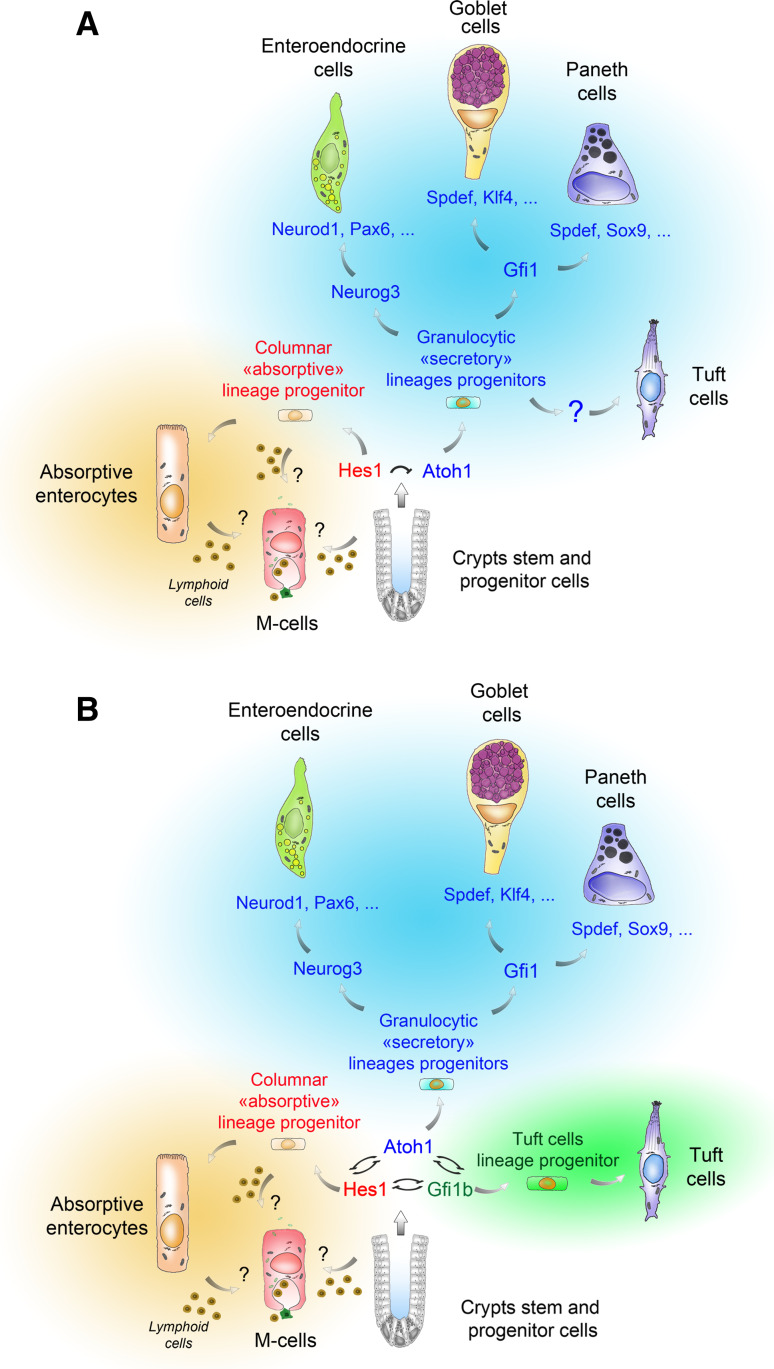
Two different schemes for tuft cell specification and differentiation, and their relationship to the other lineages. Stem cells from intestinal crypts can give rise to at least six known cell types [89]. Whether differentiation of M cells occurs directly from a progenitor or an enterocyte, and requires contacts with lymphocytes remains unclear. In model a, tuft cell specification, but not terminal differentiation, is proposed to rely on Atoh1 function. The cell fate of the stem cell progeny relies on Delta-Notch mediated lateral inhibition, leading Hes1 expressing progenitors to adopt an “absorptive” identity and Atoh1 expressing progenitors to adopt a “secretory” (granulocytic) fate. According to subsequent genetic events, Atoh1-expressing progenitors then give rise to mature enteroendocrine, goblet, Paneth or tuft cells. Neurog3 expression primes cells towards an endocrine program (Neurod1, Pax6) that results in different mature enteroendocrine subtypes. Gfi1 expression prevents ectopic Neurog3 expression in Paneth and Goblet cells. To reach a fully mature state, Paneth cells depend on the expression of Sox9 and Spdef, and goblet cells depend on that of Klf4 and Spdef. In model b, the cell fate does not rely on two but three transcription factors (Hes1, Atoh1 and Gfi1b), reciprocally antagonizing themselves. As in model a, Hes1 expression drives progenitors towards an absorptive cell identity, Atoh1 expression is essential for enteroendocrine, goblet and Paneth cell specification and survival, and Atoh1 function is not required for [64] tuft cell differentiation. The Gfi1b transcription factor, expressed in immature and terminally differentiated tuft cells, is proposed as a molecular switch towards the tuft cell lineage
References
Publication types
MeSH terms
Substances
LinkOut - more resources
Full Text Sources
Other Literature Sources
