. 2012 Jun;5(2):135-44.
doi: 10.1007/s12178-012-9123-1.

Peroneal tendoscopy

Affiliations

Peroneal tendoscopy

Antonio Marmotti et al. Curr Rev Musculoskelet Med. 2012 Jun.

Abstract

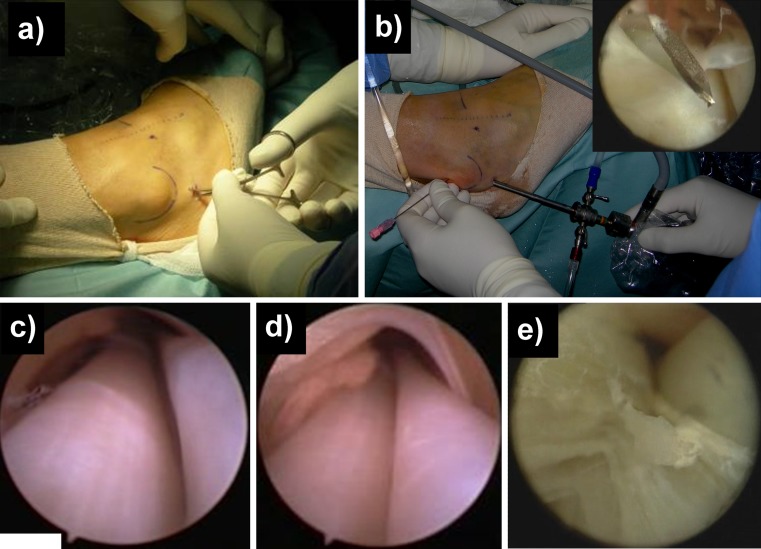
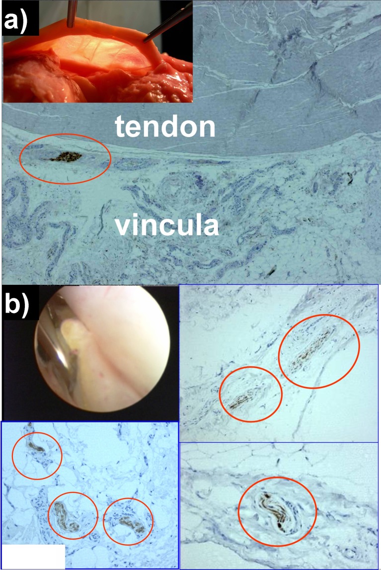
Peroneal tendoscopy is an innovative technique that allows visualization of the tendons from the myotendinous junction to the peroneal tubercle, together with adjacent anatomic structures such as the recently unveiled vincula. Through a minimally invasive approach, it is possible to diagnose and treat several disorders, such as common tenosynovitis, accessory muscles, hypertrophic bony prominences, and thickened vincula, that can cause pain and tendon catching. Surgical morbidity and postoperative pain are significantly reduced when compared with open procedures. In this paper, the main indications for peroneal tendoscopy are discussed, the available literature is reviewed, and the surgical technique is described. Advantages of this procedure and current limitations are also presented. Anatomic and histological studies were also performed in order to verify: 1) the feasibility of peroneal tendoscopy for evaluation of peroneal tendons, using cadaver specimens; 2) the presence of nervous tissue in cadaver peroneal vincula as well as in tendoscopic vincula biopsies from patients undergoing surgery for chronic lateral ankle pain.


Figures

Fig. 1
Fig. 1
a “nick and spread” technique to establish the I distal portal; b II portal placement optimized by introducing a spinal needle; c view of peroneal tendons from distal to proximal; d tenosynovitis of the peroneal tendons; e traumatic longitudinal tear of peroneus brevis
Fig. 2
Fig. 2
Immunohistochemistry for S-100 protein in vincula specimens; a cadaveric vincula sample; b vinculum biopsy harvested during tendoscopic release of a thickened vincula sample

References

    1. Christensen JC, Lanier TD. Tendoscopy of the ankle. Clin Podiatr Med Surg. 2011;28:561–570. doi: 10.1016/j.cpm.2011.05.003. - DOI - PubMed
    1. Wertheimer SJ, Weber CA, Loder BG, et al. The role of endoscopy in treatment of stenosing posterior tibial tenosynovitis. J Foot Ankle Surg. 1995;34:15–22. doi: 10.1016/S1067-2516(09)80097-5. - DOI - PubMed
    1. Dijk CN, Kort N, Scholten PE. Tendoscopy of the posterior tibial tendon. Arthroscopy. 1997;13:692–698. doi: 10.1016/S0749-8063(97)90002-5. - DOI - PubMed
    1. Sterkenburg MN, Haverkamp D, Dijk CN, Kerkhoffs GMMJ. A posterior tibial tendon skipping rope. Knee Surg Sports Traumatol Arthrosc. 2010;18:1664–1666. doi: 10.1007/s00167-010-1194-1. - DOI - PubMed
    1. Chow HT, Chan KB, Lui TH. Tendoscopic debridement for stage I posterior tibial tendon dysfunction. Knee Surg Sports Traumatol Arthrosc. 2005;13:695–698. doi: 10.1007/s00167-005-0635-8. - DOI - PubMed

LinkOut - more resources