Review
. 2012 May;90(5):495-508.
doi: 10.1007/s00109-012-0894-z. Epub 2012 Apr 14.

Emergence and evolution of the renin-angiotensin-aldosterone system

Affiliations
Review

Emergence and evolution of the renin-angiotensin-aldosterone system

David Fournier et al. J Mol Med (Berl). 2012 May.

Abstract

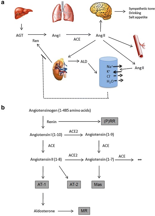
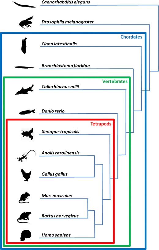
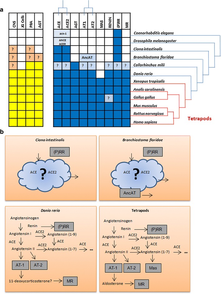
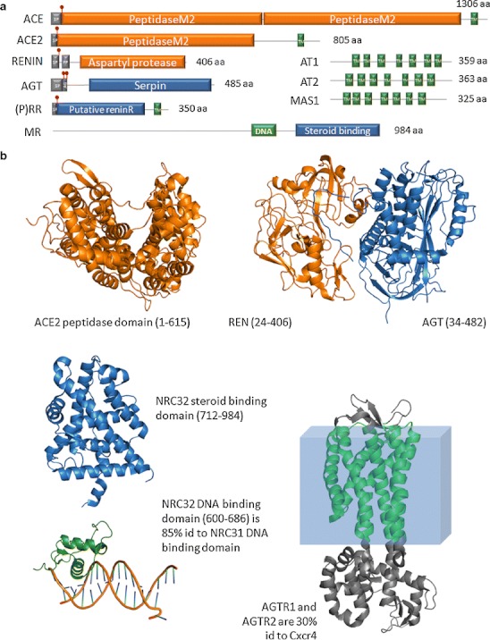
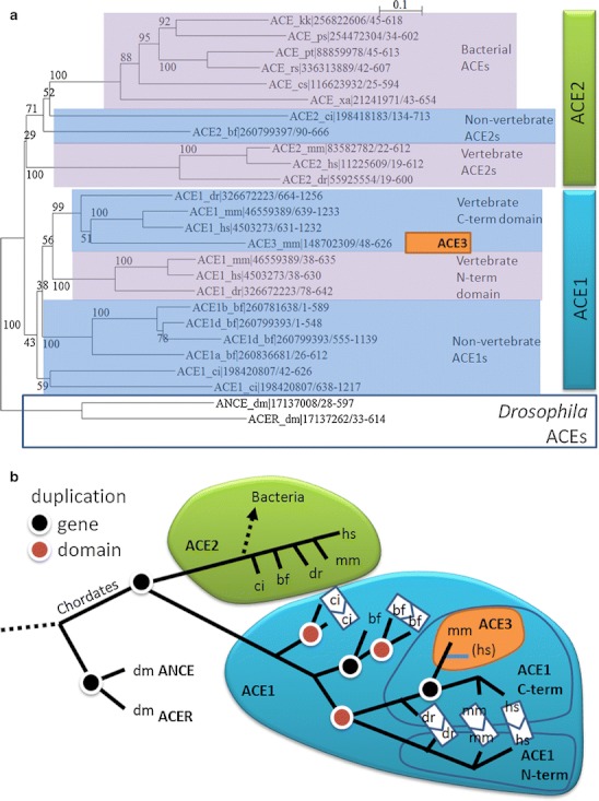
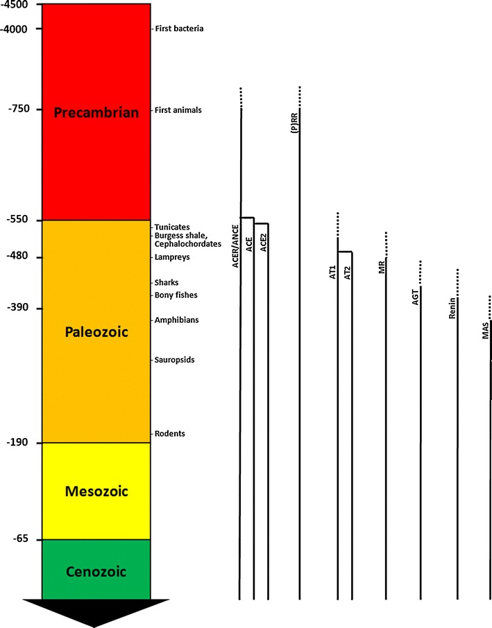
The renin-angiotensin-aldosterone system (RAAS) is not the sole, but perhaps the most important volume regulator in vertebrates. To gain insights into the function and evolution of its components, we conducted a phylogenetic analysis of its main related genes. We found that important parts of the system began to appear with primitive chordates and tunicates and that all major components were present at the divergence of bony fish, with the exception of the Mas receptor. The Mas receptor first appears after the bony-fish/tetrapod divergence. This phase of evolutionary innovation happened about 400 million years ago. We found solid evidence that angiotensinogen made its appearance in cartilage fish. The presence of several RAAS genes in organisms that lack all the components shows that these genes have had other ancestral functions outside of their current role. Our analysis underscores the utility of sequence comparisons in the study of evolution. Such analyses may provide new hypotheses as to how and why in today's population an increased activity of the RAAS frequently leads to faulty salt and volume regulation, hypertension, and cardiovascular diseases, opening up new and clinically important research areas for evolutionary medicine.


Figures

Fig. 1
Fig. 1
Scheme of the components of the hormonal RAAS. a Liver-produced angiotensin (AGT) is cleaved by renin from the kidney to the decapeptide angiotensin I (Ang I), which in turn is converted to Ang II (largely in the lung). The effector Ang II directs the adrenal gland to release aldosterone (ALD), which directs the brain to increase sympathetic tone, drinking, and salt appetite and also increases vasomotor tone. ALD, sympathetic tone, and Ang II act independently to affect NaCl reabsorption in the kidney. A reverse feedback mechanism exists. b The components involve a series of proteins (substrates, enzymes, and products) that can be defined by genomic study and followed across evolution. (P)RR prorenin receptor, ACE and ACE2 angiotensin converting enzymes 1 and 2, AT 1 and AT 2 angiotensin receptors 1 and 2, Mas Mas receptor, MR mineralocorticoid receptor. In addition to this hormonal RAAS, the system also acts locally at the tissue level, e.g., brain, adrenal gland, and heart
Fig. 2
Fig. 2
Phylogeny of the species whose sequences are studied in this review. The main groups displayed are the chordates, which comprise all species considered except Drosophila and Caenorhabditis, and display a notochord, at least at some point during their embryonic development. Vertebrates are species displaying vertebra and comprise all considered chordate species, with the exception of Ciona and Amphioxus. Finally, the tetrapods comprise all our vertebrate species, with the exception of fishes, either cartilage fishes (elephant shark, Callorhinchus Milii), bony fishes (zebrafish, Danio rerio), coelacants, or lungfishes
Fig. 3
Fig. 3
Comparison of the RAAS in multiple species. a Left panel (yellow) includes data from physiological studies: presence known or supposed of RAAS in the central nervous system (CNS), juxtaglomerular cells (JGCells), plasma renin activity (PRA), and angiotensin or angiotensin-like activity known (AGT). Note that PRA does not measure renin, but rather the conversion of AGT to Ang I; renin is not the only enzyme with this capability. Right panel (blue) shows sequence data found by BLAST inquiry. Blanks indicate instances in which the property could not be found. Question marks denote instances of uncertain or contradictory data. b Model of the stepwise emergence of the components of the RAAS based on their conservation across several taxonomic divisions. Ciona intestinalis contains the two ACEs and the prorenin receptor, but the many components missing show evidence that these three proteins have functions ancestral to the RAAS. B. floridae has an additional member, AncAT, an ancestral version of the angiotensin receptors. After a large gap, our next closer relatives whose complete genomes we know, the bony fishes, have a human-like system, with two notable differences: a possible use of a precursor of aldosterone and the absence of the Mas receptor. The tetrapods (mammals, reptiles, amphibians, birds) have the complete system, with the exception of renin, which could be missing in aves (see main text)
Fig. 4
Fig. 4
Structural features of nine human proteins relevant to the RAAS. a Domain organization of ACE, ACE2, renin, AGT, (P)RR, and MR. T transmembrane alpha-helix (TM), S signal peptide (SP), P pro-peptide (PP). Red box on angiotensinogen diagram: Ang I sequence (AG). Red symbols indicate protein cleavage sites. b Solved 3D structures of these proteins or homologs (when indicated). ACE2: peptidase domain (fragment 1-615, PDB:1R42); REN and AGT: complex of renin (blue) and AGT (orange). Note the N-terminal of AGT protruding into the renin molecule for processing (PDB:2X0B); NRC32: steroid binding domain (blue; PDB:2AA2) and DNA binding domain (green) with DNA (stick model) from 85 % identical rat glucocorticoid receptor NRC31 (PDB:3G9P); AGTR1/AGTR2 are 30 % identical to the CXCR4 chemokine receptor whose structure is shown (TM helices in green; PDB:3OE0). All protein structures are represented using the PyMOL Molecular Graphics System software (DeLano Scientific, Palo Alto, California)
Fig. 5
Fig. 5
Evolution of the ACE family. a Phylogenetic tree of the peptidase domains of selected eukaryotic and bacterial ACE homologs. The numbers at the branches indicate number of bootstrapping tests that resulted in the marked grouping: Values close to the total used (100) indicate reliable branches. The labels indicate the subfamily, a two letter abbreviation of the species name, GenPept identifier, and amino acid range. Species abbreviations of eukaryotic species are dm (Drosophila melanogaster), ci (Ciona intestinalis), bf (B. floridae), dr (Danio rerio), mm (M. musculus), and hs (H. sapiens). ACE_xa corresponds to the bacterial Xanthomonas axonopodis sequence; for the other bacterial species, please refer to the database records. Drosophila sequences contain a single domain (ANCE_dr, ; ACER_dm) and constitute an outgroup indicating that they are ancestral to chordate ACE1/ACE2. Multiple bacterial sequences (including the X. axonopodis sequence) contain a single protease domain that groups with single domain ACE2s and is not ancestral to both ACE1 and ACE2. This suggests that the bacterial sequences are a result of horizontal transfer from an ancestral chordate species. b Interpretation of the phylogenetic tree. The ACE family originated before the divergence of chordates from arthropods. Gene duplications (black dots) have expanded this family, for example, leading to the existence of ACE1 and ACE2 in chordates. Multiple events of domain duplication (red dots) have happened in the ACE1 subfamily, an important one leading to the vertebrate ACE1, which contains an N-terminal and a C-terminal catalytic domain. ACE3 is a single domain ACE, which stems from duplication of the mammalian C-terminal domain of the ACE1. This sequence seems to have evolved into a pseudogene in humans (blue line). Orthologs of vertebrate ACE2 are present in many bacterial species. Their close homology to non-vertebrate ACE2s suggests that they are the result of a single event of horizontal transfer from an ancestral non-vertebrate species. The grouping in the phylogenetic tree of the bacterial sequences analyzed here suggests that this initial event was followed by further events of horizontal transfer between bacterial species, indicating that bacterial ACEs have acquired a function that confers an evolutionary advantage to the species bearing it. Multiple sequence alignment was produced using the MUSCLE method [55] as implemented at the EBI web server. The alignment was examined, and phylogenetic trees were generated using ClustalX Version 2.1 [56] excluding positions with gaps and correcting for multiple substitutions
Fig. 6
Fig. 6
Time-line of the emergence of the RAAS. Left geological eras and a time-line (scale in millions of years). While most genes appeared in the early Paleozoic, others might have emerged earlier in the Precambrian era and were adapted for their use as part of the RAAS. ACE is one such example and might have evolved from an initial developmental function to physiological actions on volume regulation in vertebrates

References

    1. Cushman DW, Ondetti MA. Design of angiotensin converting enzyme inhibitors. Nat Med. 1999;5:1110–1113. doi: 10.1038/13423. - DOI - PubMed
    1. Ganten D, Hayduk K, Brecht HM, Boucher R, Genest J. Evidence of renin release or production in splanchnic territory. Nature. 1970;226:551–552. doi: 10.1038/226551a0. - DOI - PubMed
    1. Ganten D, Minnich JL, Granger P, Hayduk K, Brecht HM, Barbeau A, Boucher R, Genest J. Angiotensin-forming enzyme in brain tissue. Science. 1971;173:64–65. doi: 10.1126/science.173.3991.64. - DOI - PubMed
    1. Shen XZ, Xiao HD, Li P, Lin CX, Billet S, Okwan-Duodu D, Adams JW, Bernstein EA, Xu Y, Fuchs S, Bernstein KE. New insights into the role of angiotensin-converting enzyme obtained from the analysis of genetically modified mice. J Mol Med (Berl) 2008;86:679–684. doi: 10.1007/s00109-008-0325-3. - DOI - PMC - PubMed
    1. Urata H, Boehm KD, Philip A, Kinoshita A, Gabrovsek J, Bumpus FM, Husain A. Cellular localization and regional distribution of an angiotensin II-forming chymase in the heart. J Clin Invest. 1993;91:1269–1281. doi: 10.1172/JCI116325. - DOI - PMC - PubMed

LinkOut - more resources