. 2012:849:303-20.
doi: 10.1007/978-1-61779-551-0_21.

Bacterial amyloids

Affiliations

Bacterial amyloids

Yizhou Zhou et al. Methods Mol Biol. 2012.

Abstract

Many bacteria can assemble functional amyloid fibers on their cell surface. The majority of bacterial amyloids contribute to biofilm or other community behaviors where cells interact with a surface or with another cell. Bacterial amyloids, like all functional amyloids, share structural and biochemical properties with disease-associated eukaryotic amyloids. The general ability of amyloids to bind amyloid-specific dyes, such as Congo red, and their resistance to denaturation have provided useful tools for scoring and quantifying bacterial amyloid formation. Here, we present basic approaches to study bacterial amyloids by focusing on the well-studied curli amyloid fibers expressed by Enterobacteriaceae. These methods exploit the specific tinctorial and biophysical properties of amyloids. The methods described here are straightforward and can be easily applied by any modern molecular biology lab for the study of other bacterial amyloids.


Figures

Fig. 1
Fig. 1
Interbacterial complementation between an E. coli csgA mutant and a csgB mutant. (a) A schematic presentation of interbacterial complementation. A csgB mutant (the donor) secretes soluble CsgA into the media, which assembles into curli fibers on the cell surface of an adjacent csgA mutant (the recipient) expressing CsgB. (b) A csgA mutant and a csgB mutant were streaked adjacent to each other on the YESCA CR plate and incubated at 26°C for 48 h. Colonies of the csgA mutant facing to the csgB mutant stained red. (c) Western blot analysis was used to detect formation of intercellular curli fibers between an E. coli csgA mutant and a csgB mutant. Overnight cultures of csgA and csgB mutants were mixed in a 1:1 ratio and spotted onto a YESCA plate and a thin YESCA plate, respectively. Whole cell lysates and the plugs were collected for western analysis. A wild-type E. coli, a csgA and a csgB mutant were used as controls. All of the samples were treated with HFIP and probed with anti-CsgA antibody. (d) A mixed culture of E. coli csgA and csgB mutants was spotted onto YESCA plate and grown for 48 h. Curli were detected by EM. Scar bar equals to 500 nm.
Fig. 2
Fig. 2
Purified CsgA assembles into curli fibers on CsgB expressing cells. (Figure adapted from Wang et al. (29)) (a) CR staining of CsgAB+ and CsgAB overlaid with different concentrations of freshly purified CsgA. Only CsgAB+ cells with CsgA proteins stained red. (b) Negative-stain EM of CsgAB+ overlaid with freshly purified CsgA. Fibers were observed on the bacterial surface. Scar bar equals to 500 nm. (c) Negative-stain EM of CsgAB overlaid with purified CsgA. No fibers were detected. Scar bar equals to 500 nm.
Fig. 3
Fig. 3
CR staining and fluorescence quantification. (a) Use of CR staining to screen for curli mutants. Colonies of the wild-type E. coli stained red on the YESCA CR agar plate, while the curli defective mutant, csgA, remained white. Curli mutants (36) showed dark red, pink, or light pink color on the YESCA CR plate. (b–d) Measurements of the fluorescence associated with bacteria for CR (Em/Ex: 485/612 nm). BW25113 wild-type and the csgA deletion mutant strains were recovered from YESCA plates after 48 h of growth at 26°C and resuspended in 1 mL of 50 mM KPi (pH 7.2) containing 0.5 μg/mL CR or 4.5 μg/mL DAPI. After wash, serial base two dilutions are prepared and the fluorescence was measured in a 96-well plates by triplicate. As a reference, 100 μL of 50 mM KPi were used. (b) Comparison of CR fluorescence of curli producing wild-type and curli deficient csgA strains. Nonlinear (exponential fit): R 2 = 0.970 and 0.778 for BW25113 and csgA, respectively. (c) To show that the BW25113 csgA mutant was present in the same amount as the wild-type BW25113 strain, bacteria were post-labeled with the unspecific DNA 4′,6-diamidino-2-phenylindole (DAPI) fluorochrome and the DAPI fluorescence is measured (Em/Ex: 350/460 nm). Nonlinear (exponential fit): R 2 = 0.865 and 0.867 for BW25113 and csgA, respectively. (d) Quantification of CR fluorescence for bacteria prestained on YESCA CR plates. The CR fluorescence of the wild-type strain was significantly higher (around three times) compared with the BW25113 csgA mutant. ***p < 0.0001 using student’s t test. Error bars: average SEM of at least six wells per sample. The plate reader used in this determination was an Infinite 200 with the Tecan-I application and automatic optimization of the gain.
Fig. 4
Fig. 4
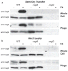
Western blot analysis of whole cell lysates and agar plugs. (a) Western blots of whole cell extracts and plugs of E. coli strains (36) that were transferred onto PVDF membranes and probed with polyclonal anti-CsgA antibodies. The csgB mutant strain did not produce curli fibers and soluble secreted CsgA was found in the agar plug. (b) Western blots of whole cell and plugs of E. coli strains (36) that were transferred onto nitrocellulose membranes and probed with polyclonal anti-CsgB antibodies. A strain lacking CsgG results in no CsgA or CsgB in whole cells or in the agar analysis.

References

    1. Cooper GJ, Willis AC, Clark A, Turner RC, Sim RB, Reid KB. Purification and characterization of a peptide from amyloid-rich pancreases of type 2 diabetic patients. Proc Natl Acad Sci USA. 1987;84:8628–8632. - PMC - PubMed
    1. Glenner GG, Wong CW. Alzheimer’s disease: initial report of the purification and characterization of a novel cerebro-vascular amyloid protein. Biochem Biophys Res Commun. 1984;120:885–890. - PubMed
    1. Prusiner SB. Molecular biology and pathogenesis of prion diseases. Trends Biochem Sci. 1996;21:482–487. - PubMed
    1. Barnhart MM, Chapman MR. Curli biogenesis and function. Annu Rev Microbiol. 2006;60:131–147. - PMC - PubMed
    1. Gebbink MF, Claessen D, Bouma B, Dijkhuizen L, Wosten HA. Amyloids--a functional coat for microorganisms. Nat Rev Microbiol. 2005;3:333–341. - PubMed

Publication types

LinkOut - more resources