Comparative Study
. 2012 Sep;470(9):2513-21.
doi: 10.1007/s11999-012-2348-x.

Does augmentation with a reinforced fascia patch improve rotator cuff repair outcomes?

Affiliations
Comparative Study

Does augmentation with a reinforced fascia patch improve rotator cuff repair outcomes?

Andrew R Baker et al. Clin Orthop Relat Res. 2012 Sep.

Abstract

Background: Scaffold devices are used to augment rotator cuff repairs in humans. While the strength of a novel poly-L-lactic acid-reinforced (human) fascia patch has been documented, it is unclear whether such patches will enhance the strength or likelihood of healing of rotator cuff repairs.

Questions/purposes: In a canine shoulder model, we asked: Do tendon repairs augmented with a reinforced fascia patch have (1) increased biomechanical properties at Time 0 and (2) less tendon retraction and increased cross-sectional area and biomechanical properties after 12 weeks of healing compared to repairs without augmentation? (3) Do the biomechanical properties of tendon repairs reach normal values by 12 weeks of healing? And (4) is the host response associated with use of the reinforced fascia patch biocompatible?

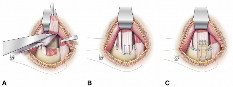
Methods: Eleven dogs underwent bilateral shoulder surgery with partial release and acute repair of the infraspinatus tendon, one shoulder with augmentation and one without augmentation. Repair retraction, cross-sectional area, biomechanical properties, and biocompatibility were assessed at 12 weeks.

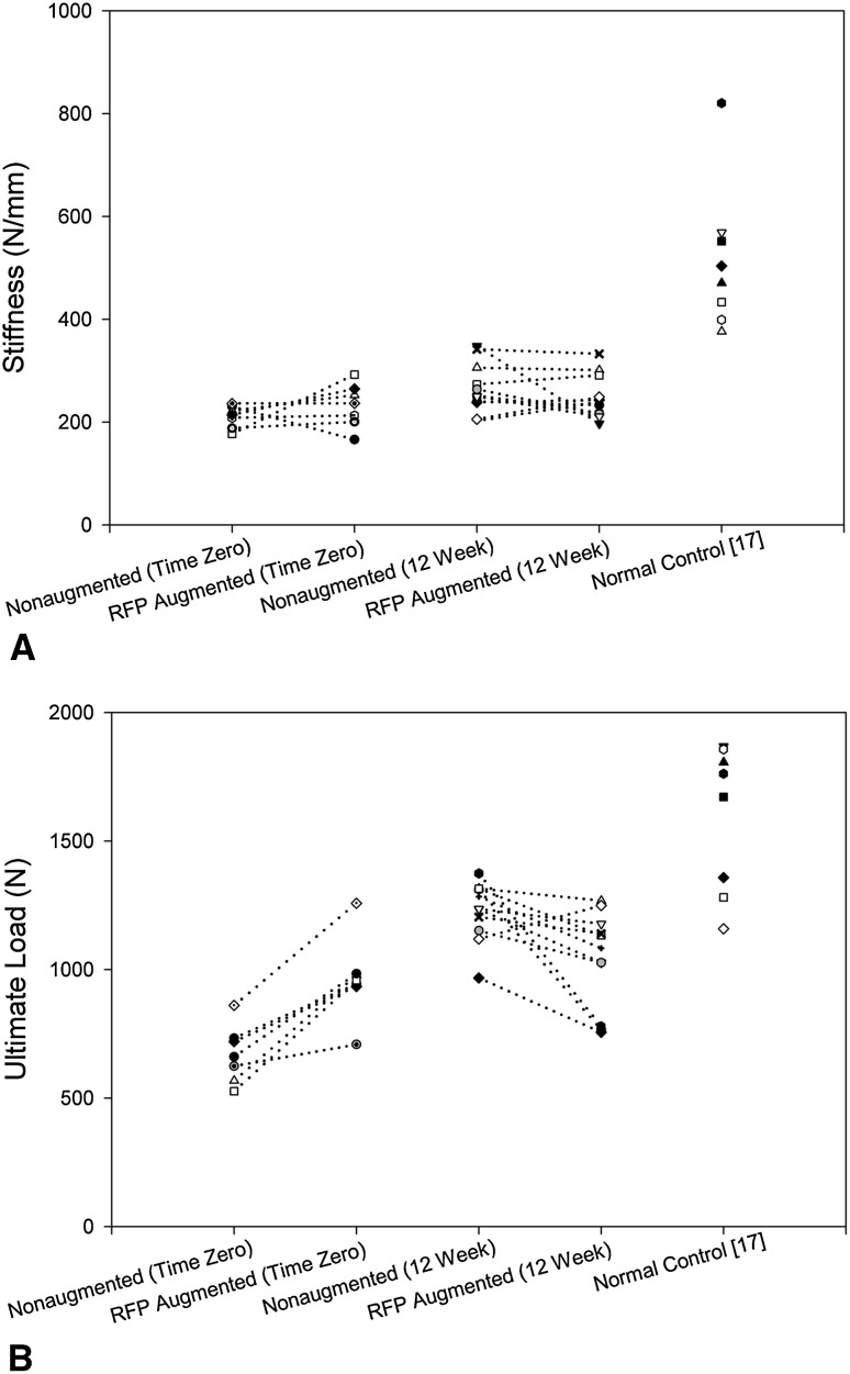
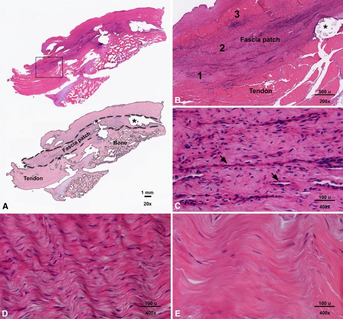
Results: At Time 0, the mean ± SD ultimate load of augmented repairs was 296 ± 130 N (46% ± 25%) more than nonaugmented repairs, with no difference in stiffness between groups. At 12 weeks, the ultimate load of augmented repairs averaged 192 ± 213 N (15% ± 16%) less than nonaugmented repairs, with no difference in stiffness between groups. At the tendon repair site at 12 weeks, the fascia patch showed a biocompatible host tissue response.

Conclusions: The biomechanical properties of repairs augmented with a reinforced fascia patch demonstrated greater ultimate load at Time 0 than nonaugmented repairs but remained essentially unchanged after 12 weeks of healing, despite improvements in the ultimate load of nonaugmented controls in the same time frame.


Figures

Fig. 1A–C
Fig. 1A–C
Diagrams illustrate bilateral rotator cuff injury and repair with and without patch augmentation in a canine model. (A) The superior 2/3 of the infraspinatus tendon was sharply detached from its insertion at the greater tuberosity, and a 1.5- × 2-cm portion of the underlying joint capsule was excised. (B) The infraspinatus tendon was immediately repaired back to its insertion using two transosseous modified Mason-Allen sutures. Bone anchors for patch fixation were included in the nonaugmented shoulders. (C) For augmentation, a fascia patch was laid over the tendon repair and attached to the tendon medially with use of three Number 0 FiberWire® Mason-Allen sutures. The device was tensioned by advancing the lateral edge approximately 2 mm laterally for osseous attachment to two customized stainless steel machine screw anchors (2.2 mm in diameter) using Number 0 FiberWire® simple sutures. Finally, the patch was tensioned superiorly and inferiorly to osseous anchors because of the absence of a soft tissue rotator cuff in this animal model. Reprinted with permission, Cleveland Clinic Center for Medical Art & Photography © 2009–2011. All rights reserved.
Fig. 2A–B
Fig. 2A–B
Graphs show the biomechanical properties of the paired nonaugmented and reinforced fascia patch (RFP) augmented repairs at Time 0 and 12 weeks. (A) There was no difference in stiffness between nonaugmented and augmented repairs at Time 0 (p = 0.219) or 12 weeks (p = 0.365). (B) At Time 0, the ultimate load of augmented repairs averaged 296 ± 130 N (46% ± 25%) more (p = 0.016) than that of nonaugmented repairs. At 12 weeks, the ultimate load of augmented repairs averaged 192 ± 213 N (15% ± 16%) less (p = 0.01) than that of nonaugmented repairs.
Fig. 3A–E
Fig. 3A–E
Images show the reinforced fascia augmented repair at 12 weeks. (A) In a representative longitudinal section of an augmented repair site (upper image), the reinforced fascia patch overlying the bone and tendon is discernable and intimately associated with surrounding host tissues (stain, hematoxylin and eosin; original magnification, ×20). In the lower image, a masked representation of the upper image outlines the fascia patch, tendon, and bone at the tendon repair site. The asterisk indicates a PLLA reinforcement fiber. (B) An inset box from (A) demonstrates neither the fascia matrix nor the PLLA reinforcing fiber appear to be resorbed in this region (stain, hematoxylin and eosin; original magnification, ×200). (C) In Region 1 from (B), the interface between the reinforced fascia patch and host tissue shows cellularized fibrous tissue with mild neovascularization (arrows; stain, hematoxylin and eosin; original magnification, ×400); (D) Region 2 from (B) demonstrates a region of high cellular density within the fascia patch (stain, hematoxylin and eosin; original magnification, ×400). (E) Region 3 from (B) demonstrates a region of low cellular density within the fascia patch (stain, hematoxylin and eosin; original magnification, ×400). Most of the cellular infiltrates within the patch were accounted for by spindle-shaped cells. There was no evidence of an inflammatory cell infiltrate, except in regions immediately adjacent to the PLLA reinforcing fiber where rare giant cells were noted.

References

    1. Accousti KJ, Flatow EL. Technical pearls on how to maximize healing of the rotator cuff. Instr Course Lect. 2007;56:3–12. - PubMed
    1. Adams JE, Zobitz ME, Reach JS, Jr, An KN, Steinmann SP. Rotator cuff repair using an acellular dermal matrix graft: an in vivo study in a canine model. Arthroscopy. 2006;22:700–709. doi: 10.1016/j.arthro.2006.03.016. - DOI - PubMed
    1. Aurora A, Mesiha M, Tan CD, Walker E, Sahoo S, Iannotti JP, McCarron JA, Derwin KA. Mechanical characterization and biocompatibility of a novel reinforced fascia patch for rotator cuff repair. J Biomed Mater Res A. 2011;99:221–230. - PubMed
    1. Badhe SP, Lawrence TM, Smith FD, Lunn PG. An assessment of porcine dermal xenograft as an augmentation graft in the treatment of extensive rotator cuff tears. J Shoulder Elbow Surg. 2008;17(1 suppl):35S–39S. doi: 10.1016/j.jse.2007.08.005. - DOI - PubMed
    1. Barber FA, Burns JP, Deutsch A, Labbe MR, Litchfield RB. A prospective, randomized evaluation of acellular human dermal matrix augmentation for arthroscopic rotator cuff repair. Arthroscopy. 2012;28:8–15. doi: 10.1016/j.arthro.2011.06.038. - DOI - PubMed

Publication types

MeSH terms