. 2012 Jun 1;287(23):19574-84.
doi: 10.1074/jbc.M111.326801. Epub 2012 Apr 23.

Induction of Bv8 expression by granulocyte colony-stimulating factor in CD11b+Gr1+ cells: key role of Stat3 signaling

Affiliations

Induction of Bv8 expression by granulocyte colony-stimulating factor in CD11b+Gr1+ cells: key role of Stat3 signaling

Xueping Qu et al. J Biol Chem. .

Abstract

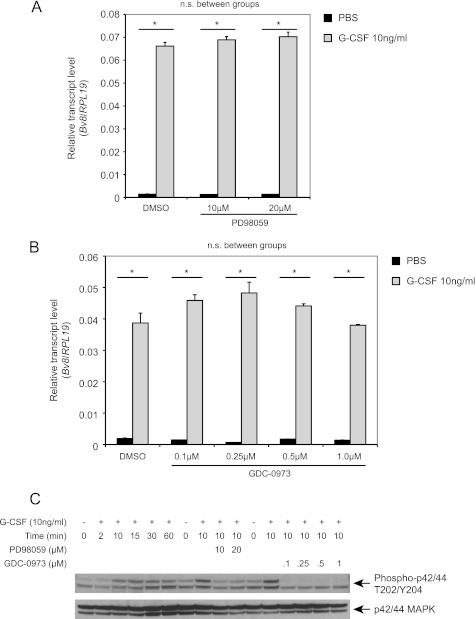
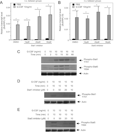
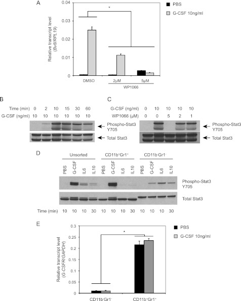
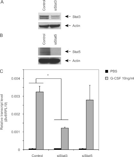
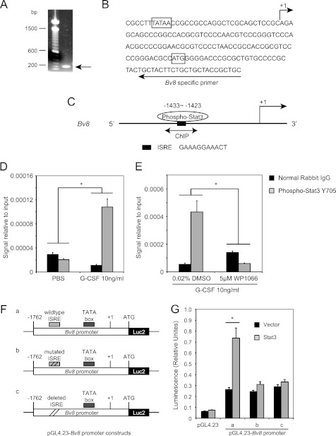
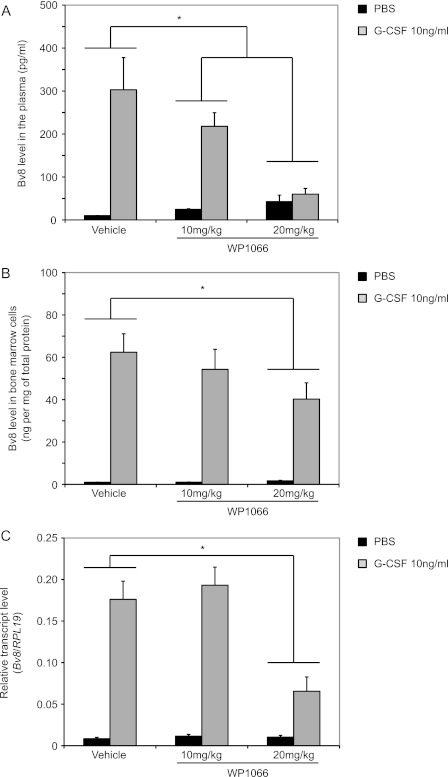
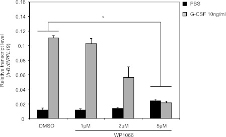
Bv8, also known as prokineticin 2, has been characterized as an important mediator of myeloid cell mobilization and myeloid cell-dependent tumor angiogenesis. Bv8 expression is dramatically enhanced by G-CSF, both in vitro and in vivo. The mechanisms involved in such up-regulation remain unknown. Using pharmacological inhibitors that interfere with multiple signaling pathways known to be activated by G-CSF, we show that signal transducer and activator of transcription 3 (Stat3) activation is required for Bv8 up-regulation in mouse bone marrow cells, whereas other Stat family members and extracellular signal-regulated kinase (ERK) activation are not involved. We further identified CD11b(+) Gr1(+) myeloid cells as the primary cell population in which Stat3 signaling is activated by G-CSF. Bv8 expression induced by G-CSF was also significantly reduced by siRNA-mediated Stat3 knockdown. Moreover, chromatin immunoprecipitation studies indicate that G-CSF significantly induces binding of phospho-Stat3 to the Bv8 promoter, which was abolished by pretreatment with the Stat3 inhibitor WP1066. Luciferase assay confirmed that the phospho-Stat3 binding site is a functional enhancer of the Bv8 promoter. The key role of Stat3 signaling in regulating G-CSF-induced Bv8 expression was further confirmed by in vivo studies. We show that the regulation of Bv8 expression in human bone marrow cells is also Stat3 signaling-dependent. Stat3 is recognized as a key regulator of inflammation-dependent tumorigenesis. We propose that such a role of Stat3 reflects at least in part its ability to regulate Bv8 expression.


Figures

FIGURE 1.
FIGURE 1.
ERK is not involved in G-CSF-induced Bv8 up-regulation. A and B, Bv8 expression in bone marrow cells incubated with the MEK-1 inhibitor PD98059 (A) or GDC-0973 (B) was assessed by TaqMan. Error bars represent S.E. Asterisks indicate significant difference between G-CSF- and PBS-treated groups (p < 0.05). n.s., not significant. C, Western blot to detect phospho-p42/44 MAPK and total p42/44 MAPK after cells had been treated with G-CSF or after pretreatment with PD98059 or GDC-0973 for 1 h followed by G-CSF for an additional 10 min. Data shown are representative of three independent experiments.
FIGURE 2.
FIGURE 2.
Stat1 and Stat5 are not involved in G-CSF induced Bv8 up-regulation. A and B, Bv8 expression analysis in bone marrow cells incubated with Stat1 (A) or Stat5 inhibitor (B). Error bars represent S.E. Asterisks indicate significant difference between G-CSF and PBS treated groups (p < 0.05). n.s., not significant. C, phospho-Stat1, phospho-Stat5, and β-actin levels after G-CSF treatment. D, phospho-Stat1 following pretreatment with Stat 1 inhibitor for 4 h. E, phospho-Stat5 after pretreatment with inhibitor for 1 h at the indicated concentrations followed by incubation with G-CSF for an additional 10 min. Data shown are representative of three independent experiments.
FIGURE 3.
FIGURE 3.
Stat3 activation is required for G-CSF induced Bv8 up-regulation. A, Bv8 expression in bone marrow cells. Error bars represent S.E. Asterisk indicates significant difference between WP1066 and control groups (p < 0.05). B–D, Western blot analysis to detect phospho-Stat3 after G-CSF treatment for the indicated time points (B), after treatment with 1, 2, or 5 μm WP1066 for 1 h followed by incubation with G-CSF for another 10 min (C), or in unsorted, CD11b+ Gr1+, and CD11b Gr1 cells after treatment with G-CSF, IL6, and IL10 as indicated (D). E, G-CSFR expression in CD11b+ Gr1+ and CD11b Gr1 cells after incubation with G-CSF or PBS for 4 h. Error bars represent S.E. Asterisk indicates significant difference in G-CSFR expression between the two cell subsets (p < 0.05). Data shown are representative of three independent experiments.
FIGURE 4.
FIGURE 4.
Stat3 siRNA inhibits G-CSF-induced Bv8 expression in bone marrow cells. Cells were transfected as indicated under “Experimental Procedures.” A and B, knockdown of Stat3 (A) or Stat5 (B) was assessed by Western blot. C, Bv8 expression was analyzed 48 h after siRNA transfection. Error bars represent S.E. Asterisk indicates significant difference between Stat3 siRNA and control siRNA treated groups (p < 0.05). Data shown are representative of three independent experiments.
FIGURE 5.
FIGURE 5.
Bv8 is a direct transcriptional target of Stat3. A, agarose gel electrophoresis of the PCR product obtained from the 5′-RACE procedure. B, sequence of the mouse Bv8 gene around the translation start codon region. The arrow above the sequence indicates the transcription start site. The arrow below the sequence indicates the mouse Bv8-specific primer used for 5′-RACE. The translation start codon ATG and the TATA box are boxed. C, schematic diagram of Stat3 binding site within the proximal promoter region of the Bv8 gene. The bidirectional arrows mark the target regions for ChIP assays. D, G-CSF significantly induces the binding of phopho-Stat3 to the putative Stat3 binding site in the Bv8 promoter region. ChIPs were performed in bone marrow cells as described under “Experimental Procedures.” Error bars represent S.E. Asterisk indicates significant difference between G-CSF- and PBS-treated groups (p < 0.05). E, binding of phospho-Stat3 to the Bv8 promoter region following G-CSF treatment was blocked by pretreatment with 5 μm WP1066. Error bars represent S.E. Asterisk indicates significant difference between WP1066- and DMSO-treated groups (p < 0.05). The amount of immunoprecipitated DNA in each sample is represented as signal relative to the total amount of input chromatin, which is equivalent to one. F, schematic diagrams of the pGL4.23-Bv8 constructs, containing wild type ISRE (a), mutated ISRE (b), and deleted ISRE (c) binding site. G, Dual-Luciferase assay to investigate the role of phospho-Stat3 ISRE site in regulating mouse Bv8 promoter activity. Error bars represent S.E. Asterisk indicates significant difference between Stat3 ORF- and control vector-transfected groups (p < 0.05).
FIGURE 6.
FIGURE 6.
WP1066 reduces the Bv8 increases in peripheral blood and bone marrow cells following in vivo administration of G-CSF. A and B, Bv8 concentrations by ELISA in plasma (A) and bone marrow cells (B). C, Bv8 expression in bone marrow cells. ELISA data were normalized to total protein concentrations. Error bars represent S.E. Asterisks indicate significant difference between WP1066- and vehicle-treated groups (p < 0.05).
FIGURE 7.
FIGURE 7.
Stat3 signaling is required for G-CSF-dependent Bv8 induction in human bone marrow cells. Human Bv8 expression by TaqMan in pooled human bone marrow cells after pretreatment with different concentrations of WP1066 for 1 h followed by incubation with 10 ng/ml human G-CSF for additional 4 h. Error bars represent S.E. Asterisk indicates significant difference between WP1066- and DMSO-treated groups (p < 0.05).

References

    1. Li M., Bullock C. M., Knauer D. J., Ehlert F. J., Zhou Q. Y. (2001) Identification of two prokineticin cDNAs: recombinant proteins potently contract gastrointestinal smooth muscle. Mol. Pharmacol. 59, 692–698 - PubMed
    1. LeCouter J., Kowalski J., Foster J., Hass P., Zhang Z., Dillard-Telm L., Frantz G., Rangell L., DeGuzman L., Keller G. A., Peale F., Gurney A., Hillan K. J., Ferrara N. (2001) Identification of an angiogenic mitogen selective for endocrine gland endothelium. Nature 412, 877–884 - PubMed
    1. Mollay C., Wechselberger C., Mignogna G., Negri L., Melchiorri P., Barra D., Kreil G. (1999) Bv8, a small protein from frog skin and its homologue from snake venom induce hyperalgesia in rats. Eur. J. Pharmacol. 374, 189–196 - PubMed
    1. Masuda Y., Takatsu Y., Terao Y., Kumano S., Ishibashi Y., Suenaga M., Abe M., Fukusumi S., Watanabe T., Shintani Y., Yamada T., Hinuma S., Inatomi N., Ohtaki T., Onda H., Fujino M. (2002) Isolation and identification of EG-VEGF/prokineticins as cognate ligands for two orphan G-protein-coupled receptors. Biochem. Biophys. Res. Commun. 293, 396–402 - PubMed
    1. Lin D. C., Bullock C. M., Ehlert F. J., Chen J. L., Tian H., Zhou Q. Y. (2002) Identification and molecular characterization of two closely related G-protein-coupled receptors activated by prokineticins/endocrine gland vascular endothelial growth factor. J. Biol. Chem. 277, 19276–19280 - PubMed

MeSH terms