

Unique MRI findings as an early predictor of osteonecrosis in pediatric acute lymphoblastic leukemia
- PMID: 22528924
- PMCID: PMC4698350
- DOI: 10.2214/AJR.11.7367
Unique MRI findings as an early predictor of osteonecrosis in pediatric acute lymphoblastic leukemia
Abstract
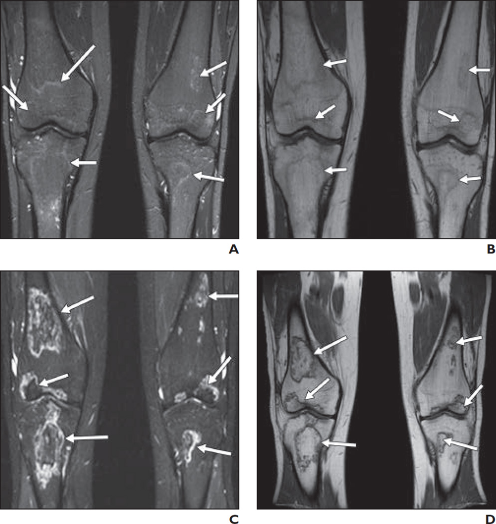
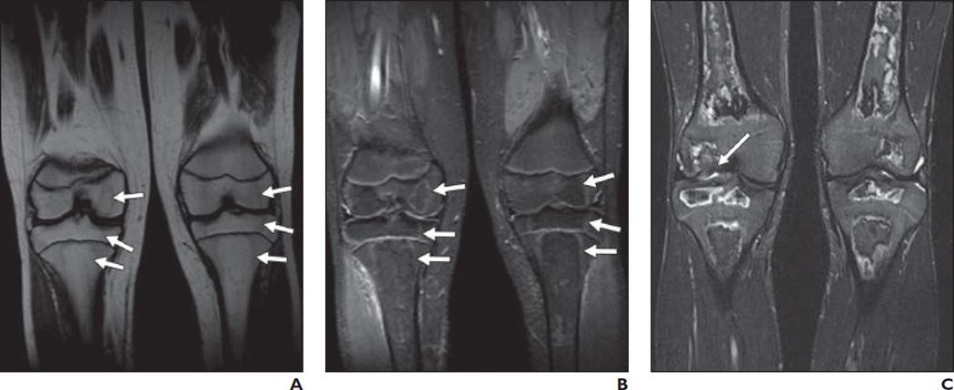
Objective: Osteonecrosis is a potential complication of glucocorticoid chemotherapy in children surviving leukemia. Early diagnosis may allow effective interventions to minimize or ameliorate joint deterioration and obviate surgical intervention. We investigated the significance of MRI signal changes that precede the currently recognized "double-line" changes, which are considered pathognomic of osteonecrosis.
Materials and methods: We retrospectively reviewed MRI scans acquired during prospective screening and follow-up of pediatric patients with leukemia for osteonecrosis.
Results: Of 481 patients, we identified 21 cases (4.3%; 12 boys; median age at leukemia diagnosis, 12.8 years) with subtle poorly defined geographically delineated MRI signal abnormalities in knees or hips, or both, that progressed over a median of 4 months (range, 1.6-18.5 months) to florid MRI signs of osteonecrosis. Articular surface collapse developed in three hips (two patients) and three knees (three patients). Three patients subsequently underwent surgical intervention (one bilateral total hip arthroplasty and one bilateral and one unilateral hip core decompression). The median duration of follow-up was 27 months (range, 1.9-90.7 months).
Conclusion: The MRI signal abnormalities described here appear to herald extensive osteonecrosis and precede the typical MRI findings of osteonecrosis previously reported in the literature.
Figures


References
-
- Ojala AE, Lanning FP, Pääkkö E, Lanning BM. Osteonecrosis in children treated for acute lymphoblastic leukemia. Med Pediatr Oncol. 1997;29:260–265. - PubMed
-
- Kaste SC, Shidler TJ, Tong X, et al. Bone mineral density and osteonecrosis in survivors of childhood allogeneic bone marrow transplantation. Bone Marrow Transplant. 2004;33:435–441. - PubMed
-
- Marker DR, Seyler TM, McGrath MS, Delanois RE, Ulrich SD, Mont MA. Treatment of early stage osteonecrosis of the femoral head. J Bone Joint Surg Am. 2008;90(suppl 4):175–187. - PubMed
-
- Mont MA, Baumgarten KM, Rifai A, Bluemke DA, Jones LC, Hungerford DS. Atraumatic osteonecrosis of the knee. J Bone Joint Surg Am. 2000;82:1279–1290. - PubMed
Publication types
MeSH terms
Substances
Grants and funding
LinkOut - more resources
Full Text Sources
Medical
