

Maternal cortisol over the course of pregnancy and subsequent child amygdala and hippocampus volumes and affective problems
- PMID: 22529357
- PMCID: PMC3356611
- DOI: 10.1073/pnas.1201295109
Maternal cortisol over the course of pregnancy and subsequent child amygdala and hippocampus volumes and affective problems
Abstract
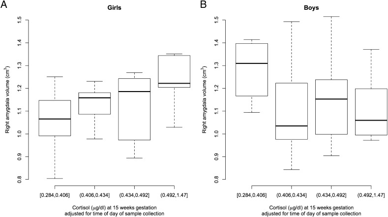
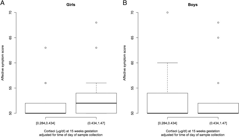
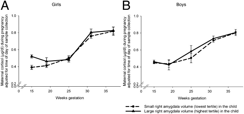
Stress-related variation in the intrauterine milieu may impact brain development and emergent function, with long-term implications in terms of susceptibility for affective disorders. Studies in animals suggest limbic regions in the developing brain are particularly sensitive to exposure to the stress hormone cortisol. However, the nature, magnitude, and time course of these effects have not yet been adequately characterized in humans. A prospective, longitudinal study was conducted in 65 normal, healthy mother-child dyads to examine the association of maternal cortisol in early, mid-, and late gestation with subsequent measures at approximately 7 y age of child amygdala and hippocampus volume and affective problems. After accounting for the effects of potential confounding pre- and postnatal factors, higher maternal cortisol levels in earlier but not later gestation was associated with a larger right amygdala volume in girls (a 1 SD increase in cortisol was associated with a 6.4% increase in right amygdala volume), but not in boys. Moreover, higher maternal cortisol levels in early gestation was associated with more affective problems in girls, and this association was mediated, in part, by amygdala volume. No association between maternal cortisol in pregnancy and child hippocampus volume was observed in either sex. The current findings represent, to the best of our knowledge, the first report linking maternal stress hormone levels in human pregnancy with subsequent child amygdala volume and affect. The results underscore the importance of the intrauterine environment and suggest the origins of neuropsychiatric disorders may have their foundations early in life.
Conflict of interest statement
The authors declare no conflict of interest.
Figures
References
-
- Geuze E, Vermetten E, Bremner JD. MR-based in vivo hippocampal volumetrics: 2. Findings in neuropsychiatric disorders. Mol Psychiatry. 2005;10:160–184. - PubMed
-
- Bellani M, Baiano M, Brambilla P. Brain anatomy of major depression II. Focus on amygdala. Epidemiol Psychiatr Sci. 2011;20:33–36. - PubMed
-
- Gluckman PD, Hanson MA. Living with the past: Evolution, development, and patterns of disease. Science. 2004;305:1733–1736. - PubMed
-
- Andersen SL. Trajectories of brain development: Point of vulnerability or window of opportunity? Neurosci Biobehav Rev. 2003;27:3–18. - PubMed
Publication types
MeSH terms
Substances
Grants and funding
LinkOut - more resources
Full Text Sources
Other Literature Sources
Medical
