Comparative Study
. 2012 May 15;109(20):E1267-76.
doi: 10.1073/pnas.1105034109. Epub 2012 Apr 23.

Dependence of Wilms tumor cells on signaling through insulin-like growth factor 1 in an orthotopic xenograft model targetable by specific receptor inhibition

Affiliations
Comparative Study

Dependence of Wilms tumor cells on signaling through insulin-like growth factor 1 in an orthotopic xenograft model targetable by specific receptor inhibition

Aleksandra Bielen et al. Proc Natl Acad Sci U S A. .

Abstract

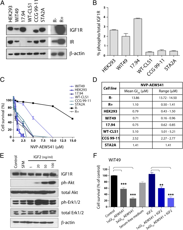
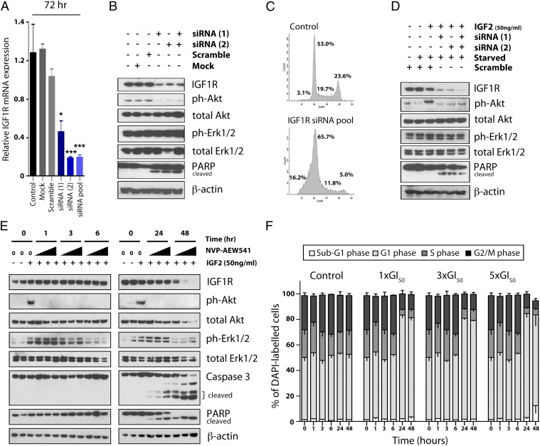
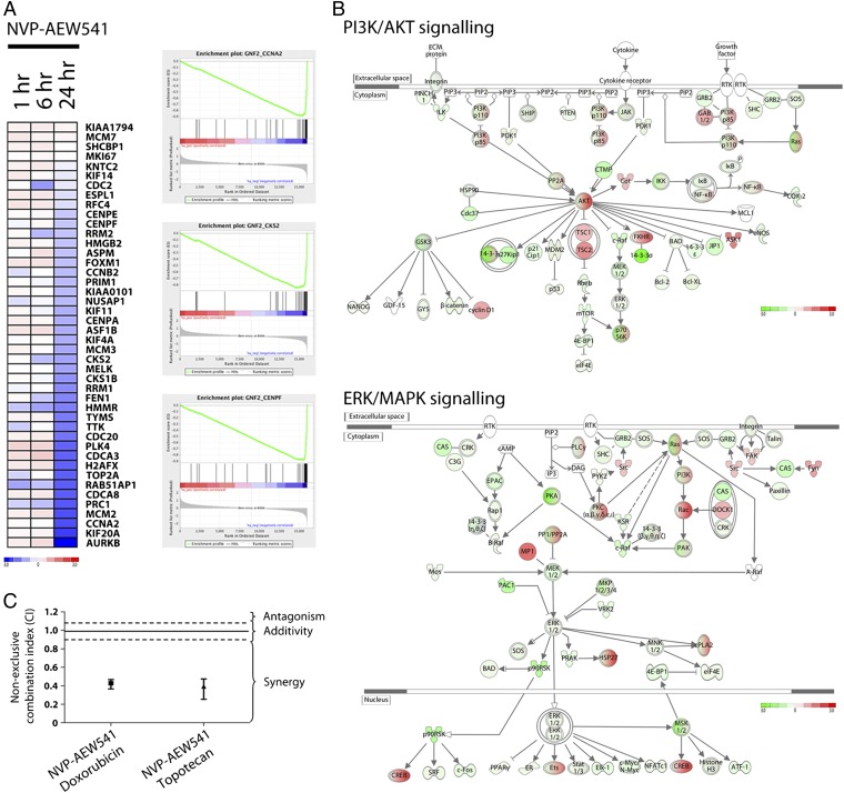
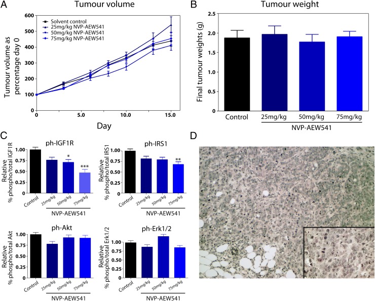
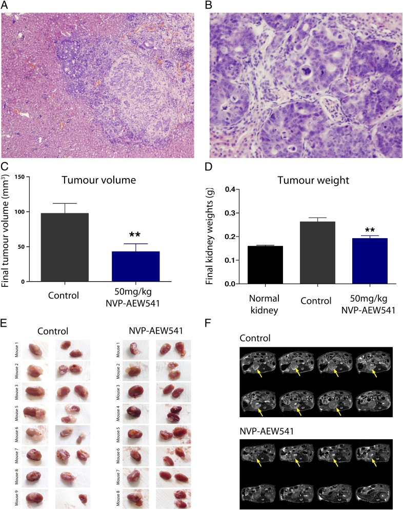
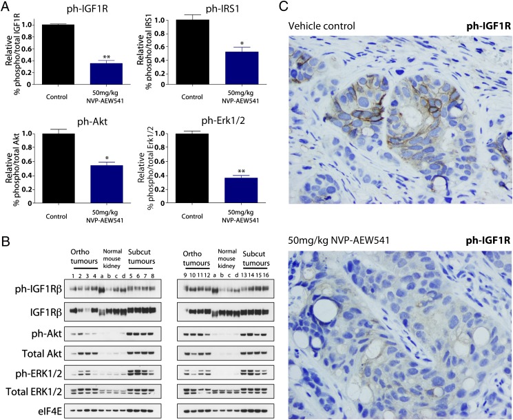
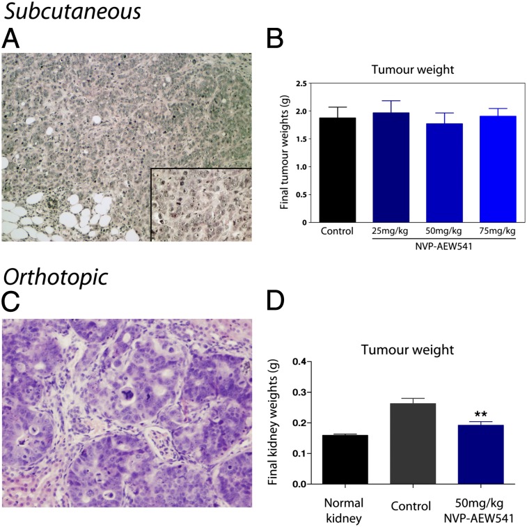
We have previously demonstrated an increased DNA copy number and expression of IGF1R to be associated with poor outcome in Wilms tumors. We have now tested whether inhibiting this receptor may be a useful therapeutic strategy by using a panel of Wilms tumor cell lines. Both genetic and pharmacological targeting resulted in inhibition of downstream signaling through PI3 and MAP kinases, G(1) cell cycle arrest, and cell death, with drug efficacy dependent on the levels of phosphorylated IGF1R. These effects were further associated with specific gene expression signatures reflecting pathway inhibition, and conferred synergistic chemosensitisation to doxorubicin and topotecan. In the in vivo setting, s.c. xenografts of WiT49 cells resembled malignant rhabdoid tumors rather than Wilms tumors. Treatment with an IGF1R inhibitor (NVP-AEW541) showed no discernable antitumor activity and no downstream pathway inactivation. By contrast, Wilms tumor cells established orthotopically within the kidney were histologically accurate and exhibited significantly elevated insulin-like growth factor-mediated signaling, and growth was significantly reduced on treatment with NVP-AEW541 in parallel with signaling pathway ablation. As a result of the paracrine effects of enhanced IGF2 expression in Wilms tumor, this disease may be acutely dependent on signaling through the IGF1 receptor, and thus treatment strategies aimed at its inhibition may be useful in the clinic. Such efficacy may be missed if only standard ectopic models are considered as a result of an imperfect recapitulation of the specific tumor microenvironment.


Conflict of interest statement

Conflict of interest statement: V.R., S.J., and F.H. are employees of Novartis Pharma.

Figures

Fig. 1.
Fig. 1.
IGF1R as a therapeutic target in Wilms tumor cell lines. (A) Western blot for IGF1R protein expression in a panel of Wilms tumor cell lines in vitro. Also included for comparison are the IGF1R-null (R−) and -overexpressing (R+) fibroblast cell lines, as well as blots for the homologous insulin receptor (IR). β-Actin is used as a loading control. (B) Quantitative measure of phosphorylated IGF1R in Wilms tumor cell lines assessed by the MSD assay. (C) Effects on cell survival of treatment with Wilms tumor cells with the IGF1R inhibitor NVP-AEW541, in comparison with the resistant R− and sensitive R+ cells. (D) GI50 values for cell lines treated with NVP-AEW541 as assessed by the MTS assay. Mean and range (in μM) for triplicate experiments are given. (E) Increased downstream signaling although PI3 and MAP kinase pathways in WiT9 cells in the presence of IGF2 assessed by Western blot analysis of phospho-Akt and phospho-Erk1/2. (F) NVP-AEW541 induces a concentration-dependent reduction in cell survival in WiT49 cells in the absence and presence of IGF2. Cells were treated with 1× and 3× GI50 concentrations of NVP-AEW541 in the absence of IGF2, and in serum-free medium including 50 ng/mL IGF2. (***P < 0.001 and **P < 0.01, t test).
Fig. 2.
Fig. 2.
Effects of genetic and pharmacological targeting of IGF1R on downstream signaling in Wilms tumor cells. (A) Relative IGF1R mRNA expression in WiT49 cells transfected with siRNA targeting the gene, as determined by quantitative RT-PCR (***P < 0.001, t test). (B) Western blots demonstrate efficient knockdown of IGF1R protein in association with diminished phospho-Akt and induced PARP cleavage in WiT49 cells transfected with IGF1R siRNA. (C) FACS analysis of siRNA-transfected WiT49 cells vs. scrambled control oligos demonstrate an accumulation of cells in G1 and sub-G1 phases in contrast to a reduction of S and G2 phases. (D) Western blots confirming the knockdown of IGF1R, reduction in phospho-Akt and phospho-Erk1/2, and induction of PARP cleavage after treatment of WiT49 cells with IGF1R siRNA in the presence of the ligand IGF2. (E) Effects on downstream signaling in WiT49 cells after treatment with NVP-AEW541 in the presence of IGF2. Cells were treated for 1, 3, 6, 24, or 48 h with 1×, 3×, or 5× GI50 compound (increase in triangle). Treatment with IGF1R inhibitor decreased phospho-Akt and phospho-Erk1/2 and induced PARP and caspase-3 cleavage in a time- and concentration-dependent manner. At 48 h, the highest concentration of compound results in a significant cell death with little protein recoverable. β-Actin is used as a loading control. (F) Effects on cell cycle in WiT49 cells treated with NVP-AEW541. An accumulation of cells in G1 and sub-G1 phases is induced by NVP-AEW541 in a time- and concentration-dependent manner. At 48 h, the highest concentration of compound results in significant cell death.
Fig. 3.
Fig. 3.
Treatment of WiT49 cells with NVP-AEW541 induces gene expression changes associated with cell cycle arrest and PI3K/MAPK down-regulation, and sensitizes the cells to chemotherapy. (A) Heat map demonstrates expression of genes associated with cell cycle progression in WiT49 cells treated with 5× GI50 of NVP-AEW541 for 1, 6, and 24 h. Genes are colored according to global, not relative, expression values (blue, down-regulated; red, up-regulated). Top-ranking gene set enrichment analysis scores for GNF2_CCNA2 [enrichment score = −0.89, P < 0.00001, false discovery rate (FDR) q < 0.00001] GNF2_CKS2 (enrichment score= −0.88, P < 0.00001, FDR q < 0.00001), and GNF2_CENPF (enrichment score = −0.85, P < 0.00001, FDR q < 0.00001) highlights coordinate down-regulation of genes in the expression neighborhood of these cell cycle control genes. (B) Top-ranking canonical pathways identified by Ingenuity Pathway Analysis after treatment of WiT49 cells with NVP-AEW541 were those associated with PI3K/AKT (P < 0.0001) and ERK/MAPK signaling (P < 0.0001). Pathway components are colored by their global gene expression levels (green, down-regulated; red, up-regulated). (C) Median effects analysis of combining NVP-AEW541 with chemotherapeutic agents in WiT49 cells. Highly synergistic interactions are seen for doxorubicin (combination index = 0.43) and topotecan (combination index = 0.39).
Fig. 4.
Fig. 4.
Treatment with NVP-AEW541 shows no efficacy in an s.c. xenograft Wilms tumor model. (A) Tumor volumes in mice implanted with WiT49 cells s.c. and treated with 25 mg/kg, 50 mg/kg, or 75 mg/kg for 15 d. Tumor volume is plotted as percentage of day 0 for all groups, with no statistically significant differences compared with vehicle-treated controls. (B) Final tumor weights for the same experiment also show no differences between treated and untreated groups. (C) Pharmacodynamic biomarkers assessed in the WiT49 s.c. model after treatment with NVP-EW541. Although a dose-dependent decrease in phospho-IGF1R and, to a lesser extent, phospho-IRS1 were observed, no significant inhibition of the downstream PI3 and MAP kinase pathways was seen. (D) Histology of the s.c. WiT49 model. The cells had prominent nucleoli and abundant slightly eosinophilic cytoplasm, with a morphological appearance more closely resembling malignant rhabdoid tumors of the kidney than classic Wilms tumors. [Original magnification of ×100 (inset, magnification of ×400).]
Fig. 5.
Fig. 5.
Antitumor activity of NVP-AEW541 in an orthotopic Wilms tumor xenograft model. (A) Low-power histological section of WiT49 cells implanted in the kidney. Tumor cells demonstrated a highly invasive phenotype, growing deep into the renal parenchyma. (Original magnification of ×100.) (B) High-power histological section of the orthotopic WiT49 tumor. Large areas with epithelial differentiation and prominent tubular formation were seen, with some cells exhibiting anaplastic features such as large cells, hyperchromatic nuclei, and atypical mitotic figures. (Original magnification of ×400.) (C) Significant reduction in tumor volume of the orthotopic WiT49 model after treatment with NVP-AEW541 (**P < 0.01, t test). (D) Significant reduction in final kidney weights of the orthotopic WiT49 model after treatment with NVP-AEW541. Tumor weights could not be directly assessed as a result of the infiltrative growth patterns observed (**P < 0.01, t test). (E) Images of the kidney in vehicle-treated control and NVP-AEW541–treated mice with orthotopic WiT49 xenografts. Extensive growth of tumor cells is observed extrarenally and deep within the kidney in the control animals, with considerably less observed in mice treated with the IGF1R inhibitor. (F) Hydrogen 1 MRI of a representative vehicle control- and NVP-AEW541–treated mouse implanted orthotopically with WiT49 cells. Tumors appear hyperintense on T2-weighted images. Considerably less tumor burden is evident in animals treated with IGF1R inhibitor compared with vehicle controls, as indicated by the yellow arrow.
Fig. 6.
Fig. 6.
Pharmacodynamic biomarkers of response of an orthotopic Wilms tumor model treated with IGF1R inhibitor. (A) MSD assay analysis of relative phospho-IGF1R, phospho-IRS1, phospho-Akt, and phospho-Erk1/2 in WiT49 cells implanted in the kidney. All markers were significantly reduced on treatment with 50 mg/kg NVP-AEW541 (**P < 0.01 and *P < 0.05, t test). (B) Western blot of constitutive levels of phospho-/total protein in WiT49 cells grown in the kidney (“ortho”) compared with the equivalent s.c. (“subcut”) model and normal mouse kidneys. (C) Representative immunohistochemistry for phospho-IGF1R in the orthotopically grown WiT49 cells demonstrates high levels in the vehicle-treated controls and significant reduction on exposure to NVP-AEW541.
Fig. P1.
Fig. P1.
Efficacy of single-agent treatment of the IGF1R small molecule inhibitor NVP-AEW541 in orthotopic, but not s.c., Wilms tumor models in vivo. WiT49 cells implanted in the flank of mice form a poorly differentiated mass of cells with little resemblance to human Wilms tumor histology (A), and which do not respond to targeted inhibition of IGF1R (B). By contrast, cells grown in the kidney invade deep into the renal parenchyma and show morphological features suggestive of anaplastic Wilms tumor (C). These tumors show a significant reduction in tumor volume and tumor weight (D) after treatment with NVP-AEW541 as a single agent.

References

    1. Pastore G, et al. Malignant renal tumours incidence and survival in European children (1978-1997): Report from the Automated Childhood Cancer Information System project. Eur J Cancer. 2006;42:2103–2114. - PubMed
    1. Kalapurakal JA, et al. Management of Wilms’ tumour: Current practice and future goals. Lancet Oncol. 2004;5:37–46. - PubMed
    1. Natrajan R, et al. Blastemal expression of type I insulin-like growth factor receptor in Wilms’ tumors is driven by increased copy number and correlates with relapse. Cancer Res. 2006;66:11148–11155. - PubMed
    1. Bjornsson HT, et al. Epigenetic specificity of loss of imprinting of the IGF2 gene in Wilms tumors. J Natl Cancer Inst. 2007;99:1270–1273. - PMC - PubMed
    1. Rainier S, et al. Relaxation of imprinted genes in human cancer. Nature. 1993;362:747–749. - PubMed

Publication types

MeSH terms