. 2012 May 8;109(19):7517-22.
doi: 10.1073/pnas.1120684109. Epub 2012 Apr 23.

Krüppel-like Factor 7 engineered for transcriptional activation promotes axon regeneration in the adult corticospinal tract

Affiliations

Krüppel-like Factor 7 engineered for transcriptional activation promotes axon regeneration in the adult corticospinal tract

Murray G Blackmore et al. Proc Natl Acad Sci U S A. .

Abstract

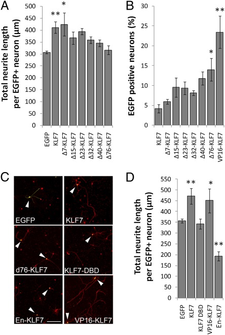
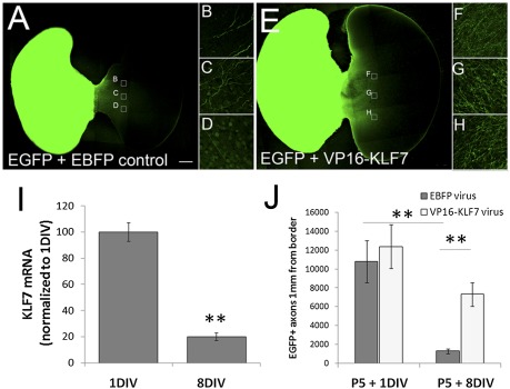
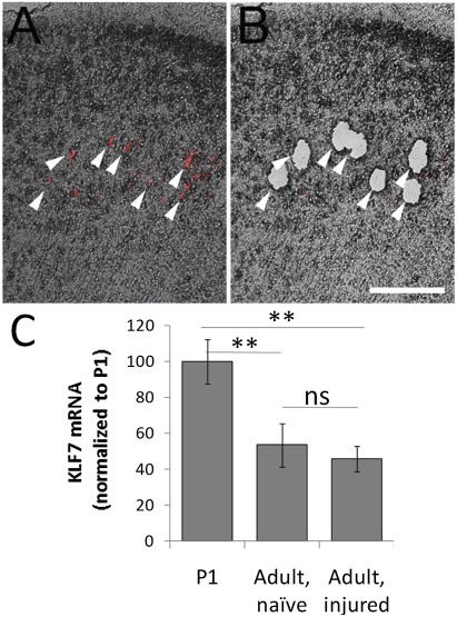
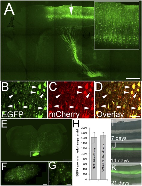
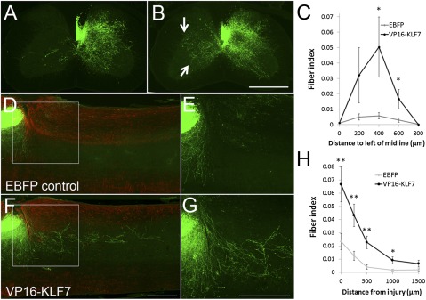
Axon regeneration in the central nervous system normally fails, in part because of a developmental decline in the intrinsic ability of CNS projection neurons to extend axons. Members of the KLF family of transcription factors regulate regenerative potential in developing CNS neurons. Expression of one family member, KLF7, is down-regulated developmentally, and overexpression of KLF7 in cortical neurons in vitro promotes axonal growth. To circumvent difficulties in achieving high neuronal expression of exogenous KLF7, we created a chimera with the VP16 transactivation domain, which displayed enhanced neuronal expression compared with the native protein while maintaining transcriptional activation and growth promotion in vitro. Overexpression of VP16-KLF7 overcame the developmental loss of regenerative ability in cortical slice cultures. Adult corticospinal tract (CST) neurons failed to up-regulate KLF7 in response to axon injury, and overexpression of VP16-KLF7 in vivo promoted both sprouting and regenerative axon growth in the CST of adult mice. These findings identify a unique means of promoting CST axon regeneration in vivo by reengineering a developmentally down-regulated, growth-promoting transcription factor.


Conflict of interest statement

Conflict of interest statement: J.L.G., V.P.L., J.L.B., and M.G.B have filed a patent pertaining to KLF-mediated neural repair.

Figures

Fig. 1.
Fig. 1.
Structure/function analysis of KLF7 identifies VP16-KLF7 as an effectively expressed growth enhancer. Cortical neurons were transfected with EGFP-tagged KLF7 constructs, cultured for 3 d, and stained for neuron-specific βIII tubulin. Neurite lengths and EGFP expression were quantified by automated image analysis (Cellomics ArrayScan VTI). (A) EGFP-KLF7 expression significantly increased neurite length, and this effect was blocked by removal of the N-terminal 76-aa acidic activation domain. (B) The percentage of neurons with detectable EGFP-KLF7 expression was increased by deletion of 76 N-terminal amino acids, and was highest in a construct in which the endogenous N-terminal domain was replaced with VP16. (C and D) Wild-type EGFP-KLF7 or EGFP-VP16-KLF7 significantly increased neurite lengths, whereas EGFP-En-KLF7 significantly decreased neurite lengths. The DNA binding domain of KLF7 alone did not affect neurite outgrowth. For each treatment, >200 individual neurons from three replicate experiments were analyzed. Error bars are SEM. **P < 0.01 vs. EGFP (A and D) or KLF7 (C), ANOVA, Dunnett’s post hoc comparison. (Scale bar: 50 μm.)
Fig. 2.
Fig. 2.
VP16-KLF7 attenuates the age-dependent decrease in axon growth in transected cortical slice cultures. Bilateral cortical slices connected by the corpus callosum were prepared from P5 rats and treated with a mixture of viral particles encoding AAV8-EGFP and either AAV8-EBFP (control) or AAV8-VP16-KLF7. After 1 or 8 DIV, the hemispheres were separated and paired with unlabeled P5 recipient tissue; axon growth was evaluated 7 d later. Slices aged 8 DIV and treated with AAV8-VP16-KLF7 (E–H) show enhanced axon growth compared with control (A–D). (I) KLF7 expression decreased ∼80% between 1 and 8 DIV. Three replicate slices from three experiments were analyzed. **P < 0.01, paired t test, mean ± SEM. (J) Axon growth from cortical slices declined about eightfold between 1 and 8 DIV. Application of VP16-KLF7 did not increase axon growth after injury at 1 DIV but increased axon growth about sixfold at 8 DIV. Error bars are SEM. **P < 0.01, ANOVA with Tukey’s post hoc comparison. Four replicate slices in two experiments were analyzed. (Scale bar: 500 μm.)
Fig. 3.
Fig. 3.
KLF7 expression in CST neurons is insensitive to cervical axotomy. CST neurons were retrogradely labeled and subjected to either sham injury or cervical dorsal transection. (A) Fluorescently labeled CST neurons in a transverse section of frontal cortex (arrows). (B) After laser capture microdissection, CST neurons were selectively gathered. (C) CST neurons were collected from adult naïve or spinally injured animals. qRT-PCR detected a 50% decrease in KLF7 expression compared with P1 cortex, independent of injury. n = 4 animals in each treatment. Error bars are SEM. **P < 0.01, ANOVA with Tukey’s post hoc comparison. (Scale bar: 100 μm.)
Fig. 4.
Fig. 4.
AAV8-EGFP identifies transduced CST neurons. (A) Sagittal brain section 4 wk after injection with AAV8-EGFP (arrow). Neuronal somata and dendrites (Inset) and descending axons are brightly labeled with EGFP. (B–D) AAV8-EGFP and AAV8-EBFP-2A-mCherry were coinjected at a 1:2 ratio. Four weeks later, nearly all EGFP+ cell bodies also express mCherry (arrowheads). (E–G) At 9 wk after cortical injection of AAV8-EGFP, the pyramidal tract is visible in horizontal sections of the medulla (box, E), and at higher magnification individual axons are clearly visible (F and G). Axon counts were sampled (boxes, F), and the total number of labeled CST axons was extrapolated from the cross-sectional area of the pyramid. (H) There was no significant difference in average axon counts between control (EBFP) and VP16KLF7-injected animals (n = 9 control, 10 VP16-KLF7 animals; mean ± SEM). (I–K) Midsagittal sections of cervical spinal cord 7, 14, and 21 d after viral injections. Error bars are SEM. A, E, F, and I–K are composites of multiple fields of view. (Scale bars: A and E, 500 μm; D, 30 μm; F, 100 μm; G, 10 μm; K, 250 μm.)
Fig. 5.
Fig. 5.
Overexpression of VP16-KLF7 promotes CST axon sprouting and regeneration in the injured spinal cord. (A and B) Transverse sections of spinal cord 4 wk after unilateral right pyramidotomy and 5 wk after coinjection of AAV8-EGFP and AAV8-EBFP (A) or AAV8-VP16-KLF7 (B) into motor cortex. Contralateral EGFP+ sprouts are abundant in VP16-KLF7–treated animals (arrow, B). Quantification (C) shows significantly elevated sprouting in VP16-KLF7 animals up to 600 μm beyond the midline. *P < 0.05, paired t test, n = 9 in each group. (D–G) Sagittal sections of cervical spinal cord 8 wk after dorsal hemisection and 9 wk after coinjection of AAV8-EGFP and AAV8-EBFP (D and E) or AAV8-VP16-KLF7 (F and G) into motor cortex. GFAP (red) shows gliosis around the injury site. EGFP+ axonal profiles (green) are visible caudal to the injury in VP16-KLF7–treated animals but not controls. (H) Quantification of EGFP+ profiles shows a significant increase up to distances of 1 mm caudal to the injury. Error bars are SEM. *P < 0.05, paired t test, n = 9 (control), 10 (VP16KLF7). A, B, D, and F are composites of multiple fields of view. (Scale bars: 500 μm.)

References

    1. Sun F, He Z. Neuronal intrinsic barriers for axon regeneration in the adult CNS. Curr Opin Neurobiol. 2010;20:510–518. - PMC - PubMed
    1. Huebner EA, Strittmatter SM. Axon regeneration in the peripheral and central nervous systems. Results Probl Cell Differ. 2009;48:339–351. - PMC - PubMed
    1. Goldberg JL, Klassen MP, Hua Y, Barres BA. Amacrine-signaled loss of intrinsic axon growth ability by retinal ganglion cells. Science. 2002;296:1860–1864. - PubMed
    1. Lemon RN, Griffiths J. Comparing the function of the corticospinal system in different species: Organizational differences for motor specialization? Muscle Nerve. 2005;32:261–279. - PubMed
    1. Cafferty WB, Duffy P, Huebner E, Strittmatter SM. MAG and OMgp synergize with Nogo-A to restrict axonal growth and neurological recovery after spinal cord trauma. J Neurosci. 2010;30:6825–6837. - PMC - PubMed

Publication types

MeSH terms

Substances

LinkOut - more resources