. 2012 May 8;109(19):7457-62.
doi: 10.1073/pnas.1200987109. Epub 2012 Apr 23.

Influenza A virus entry into cells lacking sialylated N-glycans

Affiliations

Influenza A virus entry into cells lacking sialylated N-glycans

Erik de Vries et al. Proc Natl Acad Sci U S A. .

Abstract

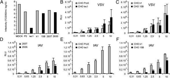
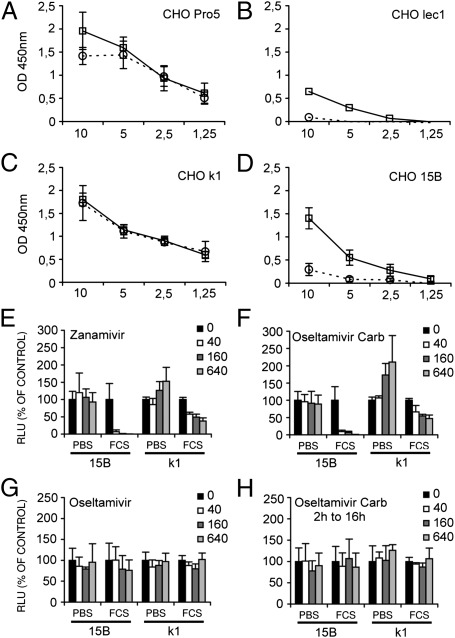
Influenza A virus (IAV) enters host cells after attachment of its hemagglutinin (HA) to surface-exposed sialic acid. Sialylated N-linked glycans have been reported to be essential for IAV entry [Chu VC, Whittaker GR (2004) Proc Natl Acad Sci USA 102:18153-18158], thereby implicating the requirement for proteinaceous receptors in IAV entry. Here we show, using different N-acetylglucosaminyl transferase 1 (GnT1)-deficient cells, that N-linked sialosides can mediate, but are not required for, entry of IAV. Entry into GnT1-deficient cells was fully dependent on sialic acid. Although macropinocytic entry appeared to be affected by the absence of sialylated N-glycans, dynamin-dependent entry was not affected at all. However, binding of HA to GnT1-deficient cells and subsequent entry of IAV were reduced by the presence of serum, which could be reversed by back-transfection of a GnT1-encoding plasmid. The inhibitory effect of serum was significantly increased by inhibition of the viral receptor-destroying enzyme neuraminidase (NA). Our results indicate that decoy receptors on soluble serum factors compete with cell surface receptors for binding to HA in the absence of sialylated N-glycans at the cell surface. This competition is particularly disturbed by the additional presence of NA inhibitors, resulting in strongly reduced IAV entry. Our results indicate that the balance between HA and NA is important not only for virion release, but also for entry into cells.


Conflict of interest statement

The authors declare no conflict of interest.

Figures

Fig. 1.
Fig. 1.
IAV infection of GnT1-deficient cells. (A) TCID50 determination after 72 h of IAV (strain WSN) incubation on the indicated WT (gray bars) or GnT1-deficient cells (black bars), demonstrating efficient infection of CHO 15B and 293S GnT1-deficient cells. (DF) Entry of increasing amounts of a luciferase-expressing IAV into WT (gray bars) or GnT1-deficient cells (black bars) as determined by luciferase expression. The x-axis indicates the amount of virus stock in inocculum; with 5 μL, ∼90% of CHO k1 or CHO Pro5 cells become infected. The y-axis represents relative light units. (B and C) Control infections by luciferase-expressing VSV.
Fig. 2.
Fig. 2.
Analysis of N-linked glycosylation in WT and GnT1-deficient CHO cell lines. (A) Western blot of HA produced in the indicated cell lines and treated with EndoH or PNGase F. HA produced in GnT1-deficient cells (Lec1 and 15B) is completely sensitive to EndoH digestion, confirming the absence of GnT1 activity in these cells. (B and C) Binding of purified soluble trimeric recombinant HA (strain WSN) to the indicated cell lines in 96-well plates. The x-axis represents HA concentration (in micrograms). NA indicates pretreatment of cells with NA. Binding was quantified by HRP-linked secondary antibodies.
Fig. 3.
Fig. 3.
Serum inhibits IAV entry into GnT1-deficient cell lines. (A–D) Entry of luciferase-expressing IAV [x-axis, virus stock (microliters); y-axis, relative light units] into the WT CHO cell lines k1 (A) and Pro5 (C) and their derivative GnT1-deficient cell lines 15B (B) and lec1 (D) was studied in the absence (black bars) or the presence (gray bars) of 10% FCS. (E and F) Entry into cells pretreated with NA (VCNA) demonstrates that entry in all cells was strictly dependent on sialic acid.
Fig. 4.
Fig. 4.
IAV entry into GnT1-deficient CHO 15B cells is strongly dependent on NA activity in the presence of serum. (A–D) Binding of recombinant soluble trimeric HA to the indicated cell lines in 96-well plates in the absence (black line) or the presence (dotted lines) of 10% serum (x-axis, HA concentration in micrograms). Binding to the parent Pro5 cells (A) or k1 cells (C) was not affected; however, binding to GnT1-deficient lec1 cells (B) and 15B cells (D) was strongly reduced in the presence of serum. (E and F) Entry of luciferase-expressing IAV (with an amount infecting ∼10% of WT CHO cells) was determined at increasing concentrations (40 nM, 160 nM, and 640 nM) of the NA inhibitors zanamivir (E) and oseltamivir carboxylate (F). The NA inhibitors strongly inhibited IAV entry into GnT1-deficient (15B) cells in the presence of serum. (G and H) Addition of the prodrug oseltamivir during entry (G) or oseltamivir carboxylate after entry (H) had no effect on luciferase expression.
Fig. 5.
Fig. 5.
(A) Pretreatment of WT CHO Pro5 cells (black bars) and CHO k1 cells (gray bars) with PNGaseF reduces entry of luciferase-expressing IAV in the presence of serum. Entry levels are plotted relative to the levels observed in nontreated cells; approximately 10% of WT CHO cells became infected at the concentration of virus used. (B and C) Transfection of GnT1-deficient 15B cells (B) and lec1 cells (C) with a GnT1 expression plasmid 24 h before inoculation fully or partially restored IAV entry into GnT1-deficient 15B and lec1 cells, respectively. (D–I) Analysis of pathway-specific entry of IAV into WT (D and E), GnT1-deficient (F and G), or GnT1-transfected (H and I) CHO cells in the presence or absence of serum. Inhibitors were 80 μM dynasore (DY), 80 μM EIPA (EI), and both DY and EI. (F, Inset) Fold inhibition of entry by 80 μM dynasore in the presence of 10% FCS in k1 cells (D), 15B cells (F), and GnT1-transfected 15B cells (H). *P < 0.01.

References

    1. Grove J, Marsh M. The cell biology of receptor-mediated virus entry. J Cell Biol. 2011;195:1071–1082. - PMC - PubMed
    1. Chu VC, Whittaker GR. Influenza virus entry and infection require host cell N-linked glycoprotein. Proc Natl Acad Sci USA. 2004;101:18153–18158. - PMC - PubMed
    1. de Vries E, et al. Dissection of the influenza A virus endocytic routes reveals macropinocytosis as an alternative entry pathway. PLoS Pathog. 2011;7:e1001329. - PMC - PubMed
    1. Sieczkarski SB, Whittaker GR. Influenza virus can enter and infect cells in the absence of clathrin-mediated endocytosis. J Virol. 2002;76:10455–10464. - PMC - PubMed
    1. Chen C, Zhuang X. Epsin 1 is a cargo-specific adaptor for the clathrin-mediated endocytosis of the influenza virus. Proc Natl Acad Sci USA. 2008;105:11790–11795. - PMC - PubMed

Publication types

MeSH terms

Substances

LinkOut - more resources