Congenital mitral valve lesions : Correlation between morphology and imaging
- PMID: 22529594
- PMCID: PMC3327011
- DOI: 10.4103/0974-2069.93703
Congenital mitral valve lesions : Correlation between morphology and imaging
Abstract
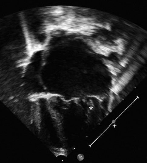
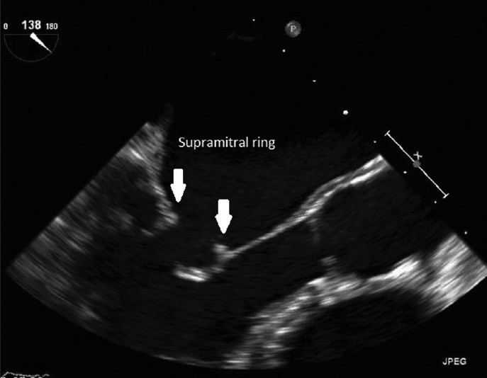
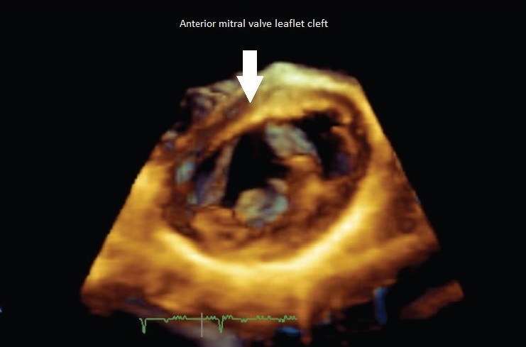
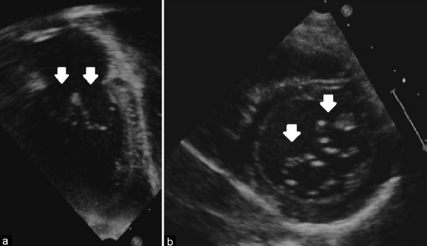
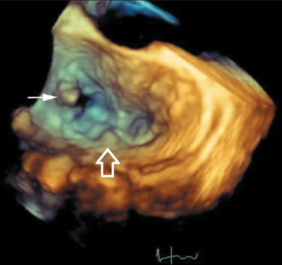
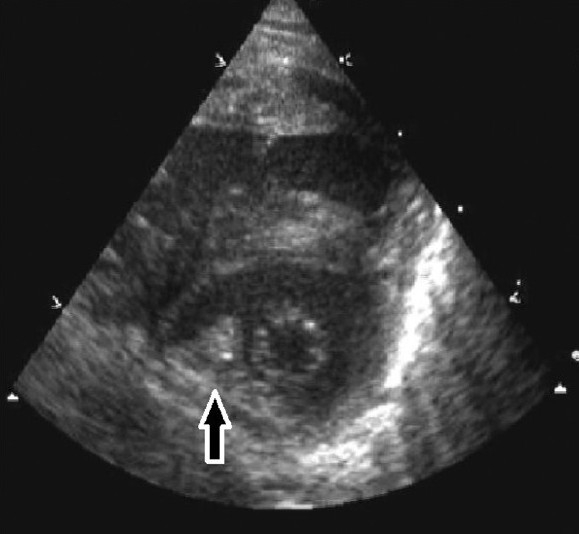
Congenital malformations of the mitral valve are often complex and affect multiple segments of the valve apparatus. They may occur in isolation or in association with other congenital heart defects. The majority of mitral valve malformations are not simply classified, and descriptive terms with historical significance (parachute, mitral, or arcade) often lack the specificity that cardiac surgeons demand as part of preoperative echocardiographic morphological assessment. This paper examines the strengths and limitations of commonly used descriptions and classification systems of congenitally malformed mitral valves. It correlates pathological, surgical, and echocardiographic findings. Finally, it makes recommendations for the systematic evaluation of the congenitally malformed mitral valve using segmental echocardiographic analysis to assist precise communication and optimal surgical management.
Keywords: Congenital; echocardiography; mitral valve.
Conflict of interest statement
Figures
References
-
- Nadas AS. Pediatric Cardiology. 3rd ed. Philadelphia: Saunders; 1972.
-
- Mitchell SC, Korones SB, Berendes HW. Congenital heart disease in 56,109 Births incidence and natural history. Circulation. 1971;43:323–32. - PubMed
-
- Hoffman JI, Kaplan S. The incidence of congenital heart disease. J Am Coll Cardiol. 2002;39:1890–900. - PubMed
-
- Celano V, Pieroni DR, Morera JA, Roland JM, Gingell RL. Two-dimensional echocardiographic examination of mitral valve abnormalities associated with coarctation of the aorta. Circulation. 1984;69:924–32. - PubMed
-
- Rosenquist GC. Congenital mitral valve disease associated with coarctation of the aorta : A0 spectrum that includes parachute deformity of the mitral valve. Circulation. 1974;49:985–93. - PubMed