Case Reports
doi: 10.4103/0974-2069.93719.
Right atrial hemangioma in the newborn: Utility of fetal imaging
Affiliations
- PMID: 22529610
- PMCID: PMC3327024
- DOI: 10.4103/0974-2069.93719
Item in Clipboard
Case Reports
Right atrial hemangioma in the newborn: Utility of fetal imaging
Ann Pediatr Cardiol.
2012 Jan.
Abstract
We present a rare primary right atrial tumor diagnosed in-utero with fetal echocardiography, and further characterized as a congenital hemangioma with magnetic resonance imaging. Surgical resection was done six days after birth. This case illustrates the complementary roles of evolving advanced imaging techniques for fetuses and infants with congenital heart disease that allows for surgery early in the neonatal period.
Keywords: Fetal echocardiography; hemangioma; right atrium.
Conflict of interest statement
Figures
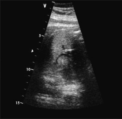
Fetal echocardiogram performed at 37-weeks gestation. Four-chamber view demonstrates nonobstructive 16 × 19 mm right atrial mass
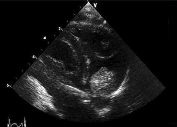
Parasternal long-axis post-natal transthoracic echocardiogram reveals a non-obstructive right atrial mass without hemodynamic compromise
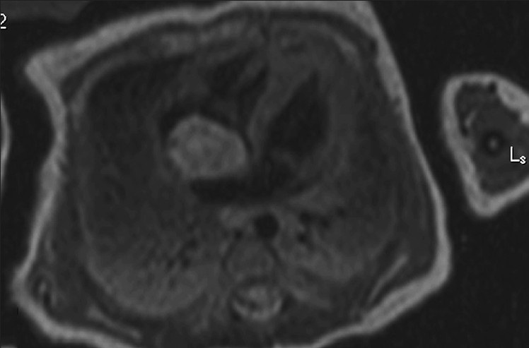
T2-weighted MRI shows a hyper-intense 1.9 × 1.4 × 1.4 cm mass arising from the infero-medial wall of the right atrium interposed between the inferior vena cava and tricuspid valve. The mass demonstrated a focal area of high attenuation in its lateral portion on pre-contrast T1-weighted imaging, suggesting a hemorrhagic or proteinaceous component
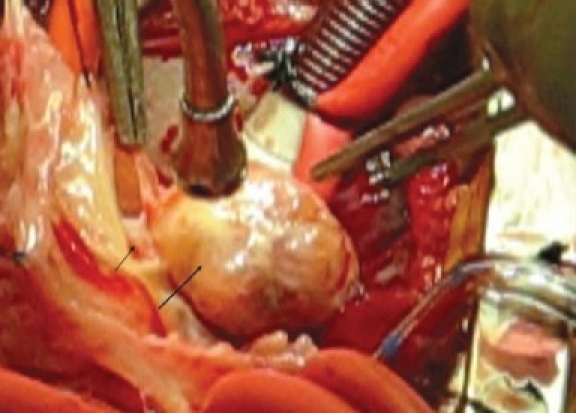
A Intra-operative image of right atrial mass viewed from the surgeon's perspective. It had attachments to the right atrial free wall and septum
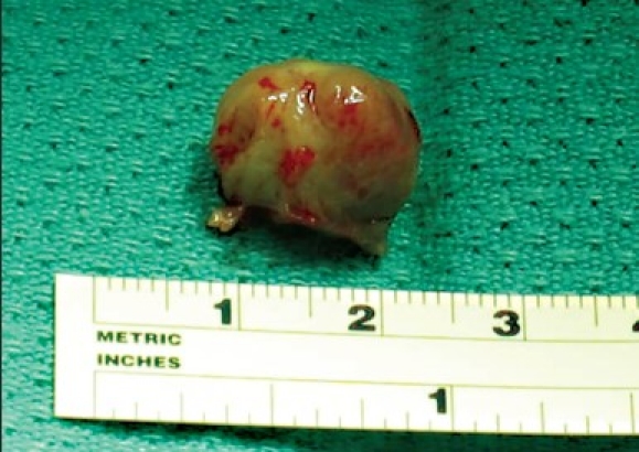
Resected right atrial mass
References
-
- Nadas AS, Ellison RC. Cardiac tumors in infancy. Am J Cardiol. 1968;21:363–6. - PubMed
-
- Hou CF, Chao A, Wang CJ, Chao AS, Hsueh C. Atrial hemangioma: A rare cause of hydrops fetalis and intrauterine fetal death. Eur J Obstet Gynecol Reprod Biol. 2007;130:271–2. - PubMed
-
- Holley DG, Martin GR, Brenner JI, Fyfe DA, Huhta JC, Kleinman CS, et al. Diagnosis and management of fetal cardiac tumors: A multicenter experience and review of published reports. J Am Coll Cardiol. 1995;26:516–20. - PubMed
-
- Ikemba CM, Eidem BW, Dimas VV, O’Day MP, Fraser CD., Jr Fetal rhabdomyoma causing postnatal critical left ventricualr outflow tract obstruction. Ann Thorac Surg. 2005;80:1529. - PubMed
-
- Isaacs H., Jr Fetal hydrops associated with tumors. Am J Perinatol. 2008;25:43–68. - PubMed