. 2012 Apr 17:3:123.
doi: 10.3389/fmicb.2012.00123. eCollection 2012.

Disassembling iron availability to phytoplankton

Affiliations

Disassembling iron availability to phytoplankton

Yeala Shaked et al. Front Microbiol. .

Abstract

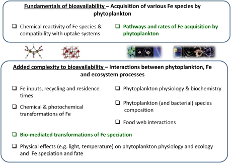
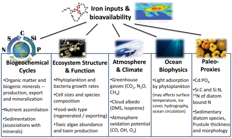
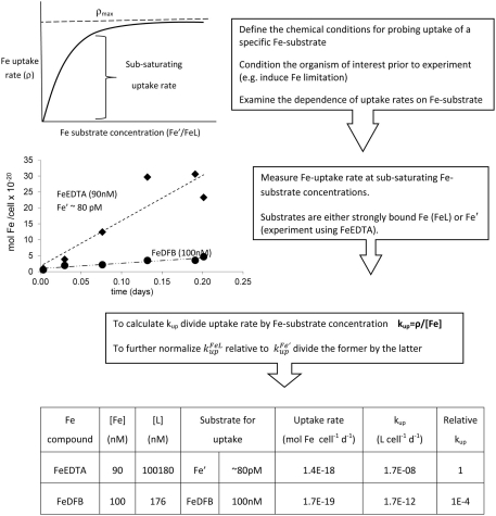
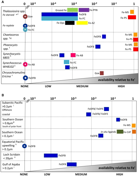
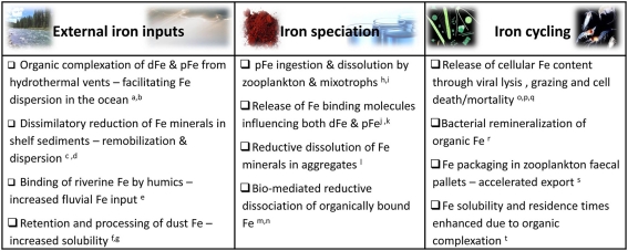
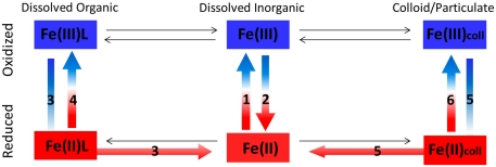
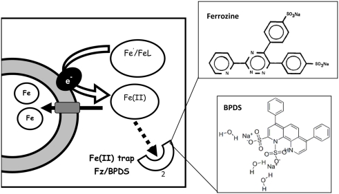
The bioavailability of iron to microorganisms and its underlying mechanisms have far reaching repercussions to many natural systems and diverse fields of research, including ocean biogeochemistry, carbon cycling and climate, harmful algal blooms, soil and plant research, bioremediation, pathogenesis, and medicine. Within the framework of ocean sciences, short supply and restricted bioavailability of Fe to phytoplankton is thought to limit primary production and curtail atmospheric CO(2) drawdown in vast ocean regions. Yet a clear-cut definition of bioavailability remains elusive, with elements of iron speciation and kinetics, phytoplankton physiology, light, temperature, and microbial interactions, to name a few, all intricately intertwined into this concept. Here, in a synthesis of published and new data, we attempt to disassemble the complex concept of iron bioavailability to phytoplankton by individually exploring some of its facets. We distinguish between the fundamentals of bioavailability - the acquisition of Fe-substrate by phytoplankton - and added levels of complexity involving interactions among organisms, iron, and ecosystem processes. We first examine how phytoplankton acquire free and organically bound iron, drawing attention to the pervasiveness of the reductive uptake pathway in both prokaryotic and eukaryotic autotrophs. Turning to acquisition rates, we propose to view the availability of various Fe-substrates to phytoplankton as a spectrum rather than an absolute "all or nothing." We then demonstrate the use of uptake rate constants to make comparisons across different studies, organisms, Fe-compounds, and environments, and for gaging the contribution of various Fe-substrates to phytoplankton growth in situ. Last, we describe the influence of aquatic microorganisms on iron chemistry and fate by way of organic complexation and bio-mediated redox transformations and examine the bioavailability of these bio-modified Fe species.

Keywords: bioavailability; biogeochemistry; iron; organic complexation; phytoplankton; redox reactions; speciation; uptake.


Figures

Figure 1
Figure 1
A conceptual diagram disassembling the multivariable concept of iron bioavailability to phytoplankton. The figure outlines the composing facets of Fe bioavailability where green text highlights topics elaborated in the paper. At the most basic level, the availability of an iron species to a phytoplankton species is determined by the rate at which it is acquired by the organism. Fe uptake rate, in turn, is a function of the uptake pathways expressed by an organism and the chemical compatibility or exchange kinetics of the Fe-substrate with the transport systems (upper box). In Sections “Fundamentals of Fe Bioavailability: Phytoplankton Fe Acquisition Systems” and “Fundamentals of Fe Bioavailability: Phytoplankton Fe Acquisition Rates” we discuss the experimental evaluation of Fe uptake rates by laboratory cultures and natural populations. In the environment, many other chemical, biological, and physical factors are important for determining Fe availability to phytoplankton, some of which are detailed in the lower box. In Section “Added Complexity to Bioavailability: Bio-Mediated Transformations of Fe Speciation” we turn to organism–Fe interactions and discuss how secretion of organic compounds and bio-mediated redox processes alter Fe speciation and influence Fe availability.
Figure 2
Figure 2
Summary of processes, systems, and research fields which are influenced by iron inputs and bioavailability (see text for details).
Figure 3
Figure 3
Prevalence of the reductive iron uptake pathway amongst phytoplankton. Listed are laboratory cultures and natural environments for which inhibition of uptake by Fe(II) binding ligands (Ferrozine/BPDS) was observed experimentally and taken to indicate the formation of an Fe(II) intermediate during iron transport. For some species, genomic and proteomic research identified various components of the reductive iron uptake pathway including ferrireductases and multicopper oxidases. See Appendix for supporting data and methodology (Tables A1 and A2 and Figures A1 and A2 in Appendix). Note on locations: The Gulf of Aqaba is located at the northern tip of the Red Sea, Loch Scridain is a sea loch located on the Atlantic coastline of the island of Mull, Scotland, and Lake Kinneret (Sea of Galilee) is a fresh water lake in the north of Israel. References: aEckhardt and Buckhout (1998), bWeger (1999), cKeshtacher et al. (1999), dAllnutt and Bonner (1987), eMiddlemiss et al. (2001), fSasaki et al. (1998), gShaked et al. (2002), hShaked et al. (2005), iJones and Morel (1988), jMaldonado and Price (2001), kSoria-Dengg and Horstmann (1995), lKranzler et al. (2011), mFujii et al. (2010), nRose et al. (2005), oMaldonado et al. (2005), pMaldonado and Price (1999), qShaked et al. (2004), rLis and Shaked (2009), Lis and Shaked, in preparation.
Figure 4
Figure 4
Schematic representation of experimental probing and calculation of the uptake rate constant – kup – exemplified with Fe-limited Emiliania huxleyi uptake data. Note that the complexing ligand L for strong ligands such as DFB should be in sufficient excess of Fe to rule out the presence of Fe′ (which would bias uptake rate since Fe′ is taken up more readily than FeL). But L:Fe should be minimal to prevent ligand from competing with cells for Fe (and thus decreasing uptake rates).
Figure 5
Figure 5
Relative scale of Fe availability established from phytoplankton uptake rates obtained for cultures (A) and natural assemblages (B). By converting experimental data to uptake-rate constants, and normalizing it further relative to Fe’, comparisons across organisms, Fe-substrates and environments are made possible (see text and Figure 4 for details). Different Fe-complexes are presented as different colors, while bar length represents variations among experiments, and circles denote no uptake. Abbreviations: Goe, goethite; DFB, desferrioxamine B; AB, Aerobactin; FC, ferrichrome; PYN, Porphyrin; MS, Monosaccharides; PS, Polysaccharides; AZ, Azotochelin; DPS, DNA binding protein from starved cells (iron storage proteins); CAT, Gallocatechin; DFE, desferrioxamine E. See Appendix for supporting data and Figure 3 for location descriptions. References: aShaked et al. (2005), bChen et al. (:1], cNodwell and Price (2001), dKustka et al. (2005), eMaldonado and Price (2001), fHassler and Schoemann (2009), gHassler et al. (2011b), hKranzler et al. (2011), iMaldonado and Price (1999), jMaldonado et al. (2005), kWells et al. (1994); lLis and Shaked (2009), Lis and Shaked, unpublished.
Figure 6
Figure 6
Selected examples of biological interactions with external iron inputs (e.g., aeolian dust deposition, sediment resuspension, fluvial, and hydrothermal Fe) and organism mediated influences on iron speciation and recycling in aquatic environments. While the microbial web in its entirety (from grazers to primary producers, viruses and heterotrophic bacteria) influences iron dynamics and availability to all community members, we focus on resultant Fe bioavailability to photosynthetic microorganisms. Abbreviations: dFe, dissolved iron; cFe, colloidal iron; pFe, particulate Fe. References: aSander and Koschinsky (2011), bWu et al. (2011), cLohan and Bruland (2008), dSevermann et al. (2010), eBatchelli et al. (2010), fBoyd et al. (2010b), gRubin et al. (2011), hBarbeau et al. (1996), iTang et al. (2011), jSato et al. (2007), kBuck et al. (2010), lBalzano et al. (2009), mMaldonado and Price (1999), nShaked et al. (2002), oHiggins et al. (2009), pStrzepek et al. (2005), qTsuda et al. (2007), rBoyd et al. (2010a), sBoyd and Ellwood (2010), tKuma et al. (1996).
Figure 7
Figure 7
Redox reactions of different Fe species in aquatic environments. See Table 2 below for details on processes 1–6 in the figure.
Figure A1
Figure A1
Trapping of ferrous iron formed during Fe′ reduction with ferrozine (Fz) or Bathophenanthrolinedisulfonic acid (BPDS). The Fe(II) trap competes with cells over ferrous iron formed during cell-mediated Fe reduction. The structures of two such traps – ferrozine and BPDS – are shown on the right top and bottom respectively. Fe(II)Fz3 and Fe(II)BPDS3 are detected spectrophotometrically at 562 and 533 nm, respectively. Additionally Fe(II)Fz3 and inhibition of Fe uptake by Fz/BPDS can be detected in radioactive experiments. Figure based on Lis and Shaked (2009).
Figure A2
Figure A2
Experimental data of iron uptake by Synechococcus WH8102 in the presence and absence of Ferrozine (Fz), on the basis of which the occurrence of the Fe reductive pathway is deduced. The uptake of iron from 90 nM 55FeDFB by iron limited Synechococcus WH8102 was inhibited in the presence of 200 μM Fz. The inhibitory effect stems from the trapping of Fe(II) formed through cell surface enzymatic reduction during Fe uptake.

References

    1. Allen M. D., Del Campo J. A., Kropat J., Merchant S. S. (2007). FEA1, FEA2, and FRE1, encoding two homologous secreted proteins and a candidate ferrireductase, are expressed coordinately with FOX1 and FTR1 in iron-deficient Chlamydomonas reinhardtii. Eukaryotic Cell 6, 1841–185210.1128/EC.00205-07 - DOI - PMC - PubMed
    1. Allnutt F. C. T., Bonner W. D. J. (1987). Evaluation of reductive release as a mechanism for iron uptake from ferrioxamine B by Chlorella vulgaris. Plant Physiol. 85, 751–75610.1104/pp.85.3.746 - DOI - PMC - PubMed
    1. Chen J.-C., Hsieh S. I., Kropat J., Merchant S. S. (2008). A ferroxidase encoded by FOX1 contributes to iron assimilation under conditions of poor iron nutrition in Chlamydomonas. Eukaryotic Cell 7, 541–54510.1128/EC.00463-07 - DOI - PMC - PubMed
    1. Chen M., Wang W. (2008). Accelerated uptake by phytoplankton of iron bound to humic acids. Aquat. Biol. 3, 155–16610.3354/ab00064 - DOI
    1. Eckhardt U., Buckhout T. J. (1998). Iron assimilation in Chlamydomonas reinhardtii involves ferric reduction and is similar to strategy I higher plants. J. Exp. Bot. 49, 1219–122610.1093/jexbot/49.324.1219 - DOI

LinkOut - more resources