. 2012;7(4):e35206.
doi: 10.1371/journal.pone.0035206. Epub 2012 Apr 17.

Mycobacterium tuberculosis Rv3586 (DacA) is a diadenylate cyclase that converts ATP or ADP into c-di-AMP

Affiliations

Mycobacterium tuberculosis Rv3586 (DacA) is a diadenylate cyclase that converts ATP or ADP into c-di-AMP

Yinlan Bai et al. PLoS One. 2012.

Abstract

Cyclic diguanosine monophosphate (c-di-GMP) and cyclic diadenosine monophosphate (c-di-AMP) are recently identified signaling molecules. c-di-GMP has been shown to play important roles in bacterial pathogenesis, whereas information about c-di-AMP remains very limited. Mycobacterium tuberculosis Rv3586 (DacA), which is an ortholog of Bacillus subtilis DisA, is a putative diadenylate cyclase. In this study, we determined the enzymatic activity of DacA in vitro using high-performance liquid chromatography (HPLC), mass spectrometry (MS) and thin layer chromatography (TLC). Our results showed that DacA was mainly a diadenylate cyclase, which resembles DisA. In addition, DacA also exhibited residual ATPase and ADPase in vitro. Among the potential substrates tested, DacA was able to utilize both ATP and ADP, but not AMP, pApA, c-di-AMP or GTP. By using gel filtration and analytical ultracentrifugation, we further demonstrated that DacA existed as an octamer, with the N-terminal domain contributing to tetramerization and the C-terminal domain providing additional dimerization. Both the N-terminal and the C-terminal domains were essential for the DacA's enzymatically active conformation. The diadenylate cyclase activity of DacA was dependent on divalent metal ions such as Mg(2+), Mn(2+) or Co(2+). DacA was more active at a basic pH rather than at an acidic pH. The conserved RHR motif in DacA was essential for interacting with ATP, and mutation of this motif to AAA completely abolished DacA's diadenylate cyclase activity. These results provide the molecular basis for designating DacA as a diadenylate cyclase. Our future studies will explore the biological function of this enzyme in M. tuberculosis.


Conflict of interest statement

Competing Interests: The authors have declared that no competing interests exist.

Figures

Figure 1
Figure 1. Purification and oligomerization of DacA.
(A) SDS-PAGE of purified DacA. Lane M, EZ-Run Pre-stained Rec Protein Ladder (Fischer Scientific); lane 1, purified DacA. (B) Gel filtration chromatograph of DacA. The molecular weights (in kDa) and the retention volumes of the standards are indicated on the top. (C) Analytical ultracentrifugation of DacA. Molecular mass of DacA was estimated using the c(M) method.
Figure 2
Figure 2. Determination of DacA's activities using HPLC and LC-MS.
(A) Analysis of the products from reaction of ATP with DacA using HPLC. Reaction of ATP with DisA was included as a positive control. The reactions were carried out as described in the Methods. ATP, c-di-AMP, ADP, AMP and pApA standards were also analyzed under the same conditions. (B and C) LC/UV/MS profiles of the products formed by DacA with ATP. The products were detected by monitoring EMS at mass range from 100 to 1000 amu (B) or monitored by UV absorption at 254 nm (C).
Figure 3
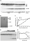
Figure 3. Determination of DacA's activities using TLC.
(A) Separation of nucleotides generated from [α-33P]ATP by DacA and DisA. The positions of ATP, ADP, AMP and c-di-AMP are indicated based on the Rf of each standard analyzed in panel B under the same conditions. (B) Separation of nucleotide standards using TLC. Spots 1–5 are ATP, ADP, AMP, c-di-AMP and pApA, respectively. (C) Quantitation of c-di-AMP production. The relative intensity of c-di-AMP generated by DacA or DisA at various time points as in panel A was analyzed using the ImageQuant software. Data shown are representative of two repeat experiments. (D) Production of c-di-AMP with various concentrations of DacA at 30 min of incubation. Reactions contain 2-fold serial diluted DacA protein as indicated on the top of the TLC graph (in log2 µg). “N” indicates a control with no protein, and “Ctl” contains 1 µg DisA as a positive control. “0” equals 1 µg of protein. (E) Quantitation of ATP depletion and c-di-AMP production by DacA from panel D. Data shown are representative of two repeat experiments.
Figure 4
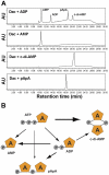
Figure 4. Catalytic activities of DacA with different nucleotides.
(A) Reaction of DacA with ADP, AMP, c-di-AMP or pApA. Samples were separated by HPLC. The peaks in “DacA+ADP” are labeled according to the retention time of each standard as shown in Fig. 2A. (B) Reactions catalyzed by DacA using ATP as a substrate, based on the results shown in Fig. 2 and Fig. 4A. “A” stands for adenosine, and “P” stands for phosphate. The thickness of arrows denotes priority of reaction, and the thickest arrow shows the major catalytic reaction.
Figure 5
Figure 5. Effect of divalent metal ions and pH on DacA's activities.
(A and B) Effect of metal ions on c-di-AMP production catalyzed by DacA in the presence of 2 mM ATP (A) or 0.5 mM ADP (B). (C and D) Effect of pH on c-di-AMP production catalyzed by DacA in the presence of 2 mM ATP (C) or 0.5 mM ADP (D). Note that less ADP was used in the reactions compared with ATP, and thus the arbitrary units between reactions with ATP and ADP are not directly comparable.
Figure 6
Figure 6. Function of the N-terminal and the C-terminal domains of DacA in oligomerization and enzymatic activity.
(A) Schematic representation of the primary structures of DacA, DacA1–287 and DacA140–358, as indicated with black lines. DacA1–287 lacks the C-terminal HhH domain, while DacA140–358 lacks the N-terminal Dac domain. (B) SDS-PAGE of purified DacA1–287 and DacA140–358. Lane M, MW marker; lanes 1 and 2 are purified DacA1–287 and DacA140–358, respectively. (C) Analytical ultracentrifugation of DacA1–287 and DacA140–358. (D) Cross-linking of DacA140–358 with glutaraldehyde. Lane M, MW marker; lane 1, untreated DacA140–358; and lane 2, glutaraldehyde-treated DacA140–358. Lanes 1 and 2 were analyzed using Western blot with the anti-DacA antibody. (E) ATP binding by DacA, DacA1–287 and DacA140–358. Proteins, either in the presence (+) or absence (−) of ATP, were separated by electrophoresis with a native gel and stained with Coomassie Brilliant Blue. (F) Enzymatic activity of 10 µM DacA1–287 and DacA140–358 analyzed using HPLC.
Figure 7
Figure 7. Function of the DGA and RHR motifs in DacA.
(A) Partial sequence alignment of M. tuberculosis DacA and T maritima DisA showing the conserved DGA and RHR motifs. Identical residues between the two proteins are highlighted in yellow blocks. (B) Potential contact of DGA and RHR motifs with ATP generated from T. maritima DisA using the Cn3D software. The three amino acids highlighted in yellow represent either DGA or RHR, as indicated. (C) SDS-PAGE of purified DacA, DacARHR and DacAG73A. Lane M, MW marker; lanes 1–3 are purified DacA, DacARHR and DacAG73A, respectively. (D) ATP binding by DacARHR and DacAG73A. Proteins either in the presence (+) or absence (−) of ATP were separated by electrophoresis with a native gel and stained with Coomassie Brilliant Blue. (E) Analysis of diadenylate cyclase activity of 10 µM DacAG73A and DacARHR in the presence of ATP using HPLC. (F) Structural modeling of DacA and its derivative polypeptides. Three domains of DacA from the N-terminus to the C-terminus are colored in green, blue and orange, respectively. A red star represents one molecule of ATP. A yellow line in DacARHR indicates the mutation of RHR motif.

References

    1. Aziz MA, Wright A. The World Health Organization/International Union Against Tuberculosis and Lung Disease Global Project on Surveillance for Anti-Tuberculosis Drug Resistance: a model for other infectious diseases. Clin Infect Dis. 2005;41(Suppl 4):S258–262. - PubMed
    1. Corbett EL, Watt CJ, Walker N, Maher D, Williams BG, et al. The growing burden of tuberculosis: global trends and interactions with the HIV epidemic. Arch Intern Med. 2003;163:1009–1021. - PubMed
    1. Espinal MA. The global situation of MDR-TB. Tuberculosis (Edinb) 2003;83:44–51. - PubMed
    1. Fine PE. Variation in protection by BCG: implications of and for heterologous immunity. Lancet. 1995;346:1339–1345. - PubMed
    1. Gomelsky M. cAMP, c-di-GMP, c-di-AMP and now cGMP: bacteria use them all! Mol Microbiol. 2011;79:562–565. - PMC - PubMed

Publication types

MeSH terms

LinkOut - more resources