. 2012;7(4):e35213.
doi: 10.1371/journal.pone.0035213. Epub 2012 Apr 18.

Propylthiouracil is teratogenic in murine embryos

Affiliations

Propylthiouracil is teratogenic in murine embryos

Valeria C Benavides et al. PLoS One. 2012.

Abstract

Background: Hyperthyroidism during pregnancy is treated with the antithyroid drugs (ATD) propylthiouracil (PTU) and methimazole (MMI). PTU currently is recommended as the drug of choice during early pregnancy. Yet, despite widespread ATD use in pregnancy, formal studies of ATD teratogenic effects have not been performed.

Methods: We examined the teratogenic effects of PTU and MMI during embryogenesis in mice. To span different periods of embryogenesis, dams were treated with compounds or vehicle daily from embryonic day (E) 7.5 to 9.5 or from E3.5 to E7.5. Embryos were examined for gross malformations at E10.5 or E18.5 followed by histological and micro-CT analysis. Influences of PTU on gene expression levels were examined by RNA microarray analysis.

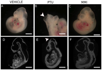
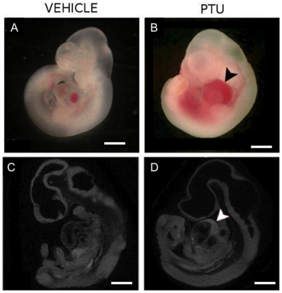
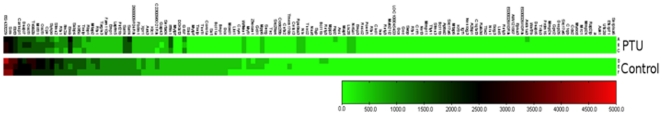
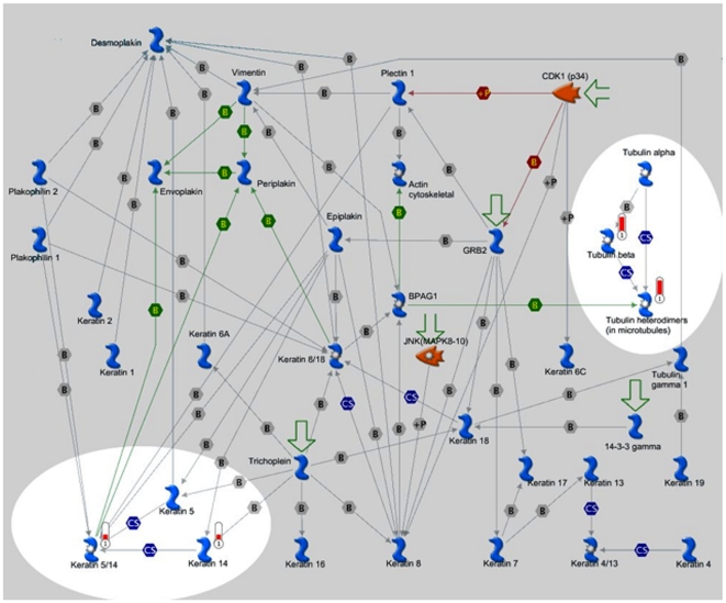
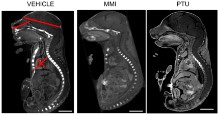
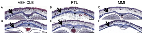
Results: When dams were treated from E7.5 to E9.5 with PTU, neural tube and cardiac abnormalities were observed at E10.5. Cranial neural tube defects were significantly more common among the PTU-exposed embryos than those exposed to MMI or vehicle. Blood in the pericardial sac, which is a feature indicative of abnormal cardiac function and/or abnormal vasculature, was observed more frequently in PTU-treated than MMI-treated or vehicle-treated embryos. Following PTU treatment, a total of 134 differentially expressed genes were identified. Disrupted genetic pathways were those associated with cytoskeleton remodeling and keratin filaments. At E 18.5, no gross malformations were evident in either ATD group, but the number of viable PTU embryos per dam at E18.5 was significantly lower from those at E10.5, indicating loss of malformed embryos. These data show that PTU exposure during embryogenesis is associated with delayed neural tube closure and cardiac abnormalities. In contrast, we did not observe structural or cardiac defects associated with MMI exposure except at the higher dose. We find that PTU exposure during embryogenesis is associated with fetal loss. These observations suggest that PTU has teratogenic potential.


Conflict of interest statement

Competing Interests: The authors have declared that no competing interests exist.

Figures

Figure 1
Figure 1. PTU exposure during embryogenesis induces cranial defects.
Representative images of E10.5 embryos treated with (A, D) vehicle, (B, E) PTU (100 mg/kg) or (C, F) MMI (20 mg/kg) illustrate un-fused cephalic neural folds in PTU-treated embryos (arrowhead). (D–F) MicroCT scans showing sagittal 3D images of E10.5 whole embryos with arrowhead identifying unfused neural folds defect. Scale bars = 0.5 mm.
Figure 2
Figure 2. PTU exposure during embryogenesis induces blood in the pericardium.
Representative images of E10.5 embryos treated with (A, C) vehicle and (B, D) PTU (100 mg/kg) showing blood in the pericardial sac of PTU-treated embryo (arrowhead). (C, D) MicroCT scans showing sagittal 3D images of whole embryos. Scale bars = 0.5 mm.
Figure 3
Figure 3. Heat map showing the differential gene expression of 139 genes between PTU and vehicle treated groups.
Heat map generated in GenomeStudio compares the average detection signals of genes between PTU and control groups. Total RNA was collected at E10.5 from whole-embryos treated between E7.5–E9.5,. Three PTU and three control RNA samples were tested and genes with a significant p-value (p<0.001) were displayed. Scale bar indicates detection signal.
Figure 4
Figure 4. Expression of Genes in the Cytoskeleton remodeling and keratin filament pathways were altered by PTU treatment.
Cytoskeleton structure in most eukaryotic cells consists of three distinct interconnected, filament systems: Actin filaments, Microtubules and intermediate filaments. Cell assembly is integrated by the network of intermediate filaments (IFs) and by their interactions with other cytoskeleton structural elements defining cytoarchitecture and cytodynamics. The IF network is critically involved in cell shape control and imparts intracellular mechanical strength. The experimental data are visualized on the map in red (increased expression or up-regulation) histogram. The height of the histogram corresponds to the relative expression value for a particular gene/protein.
Figure 5
Figure 5. PTU exposure during embryogenesis and effects in E18.5 embryos.
Embryos treated in utero from E7.5–E9.5 were analyzed at E18.5. Representative MicroCT scans showing sagittal 3D images of whole embryos indicate no differences between the treatment groups of vehicle, PTU, and MMI. Left panel shows location of morphometic measurements (a. Snout: upper lip to soft palate. b. Fronto-occipital: frontal to occipital lobes. c. Aorta-apex diameter: aortic valve to left ventricular. d. Biventricular diameter: right to left ventricle). Scale bars = 5 mm.
Figure 6
Figure 6. Representative histopathology of E18.5 calveria from vehicle, PTU and MMI treated mice.
Histopathologic analysis of coronal sections through the head of E18.5 embryos from dams treated with (A, D) vehicle, (B, E) PTU (100 mg/kg), or (C, F) MMI (20 mg/kg) from E7.5 to E9.5 did not reveal any significant morphologic changes in the bones of the calveria (arrowheads) or the connective tissue stroma within the (A, B, C) frontal or (D, E, F) parietal sutures when compared to the skulls of control embryos. Representative sections from (A, B, C) rostral dorsal frontal bone at the level of the nasal cortex, and (D, E, F) caudal dorsal parietal at the level of the ears. Scale bars = 200 µm. Masson's Trichrome stained sections are shown.

References

    1. Weetman A. Grave's disease 1835–2002. Horm Res. 2003;59(Suppl 1):114–118. - PubMed
    1. Cooper D. Antithyroid drugs. N Engl J Med. 2005;352:905–917. - PubMed
    1. Rivkees SA. Pediatric Graves' Disease: Controversies in Management. Horm Res Paediatr. 2010;74(5):305–311. - PubMed
    1. Patil-Sisodia K, Mestman J. Graves hyperthyroidism and pregnancy: a clinical update. Endocr Pract. 2010;16:118–129. - PubMed
    1. Martin J, Hamilton B, Sutton P, Ventura S, Menacker F, et al. National Vital Statistics Reports. 7. Vol. 57. Hyattsville, MD: Centers for Disease Control and Prevention; 2009. Births: Final Data for 2006.

Publication types

MeSH terms