Clinical Trial
. 2012;7(4):e35241.
doi: 10.1371/journal.pone.0035241. Epub 2012 Apr 17.

Iron accumulation in deep cortical layers accounts for MRI signal abnormalities in ALS: correlating 7 tesla MRI and pathology

Affiliations
Clinical Trial

Iron accumulation in deep cortical layers accounts for MRI signal abnormalities in ALS: correlating 7 tesla MRI and pathology

Justin Y Kwan et al. PLoS One. 2012.

Abstract

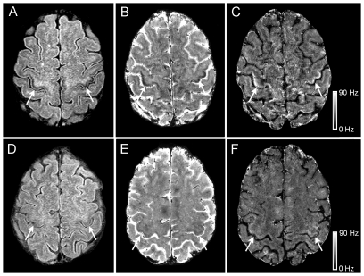
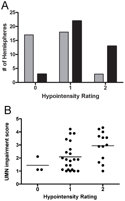
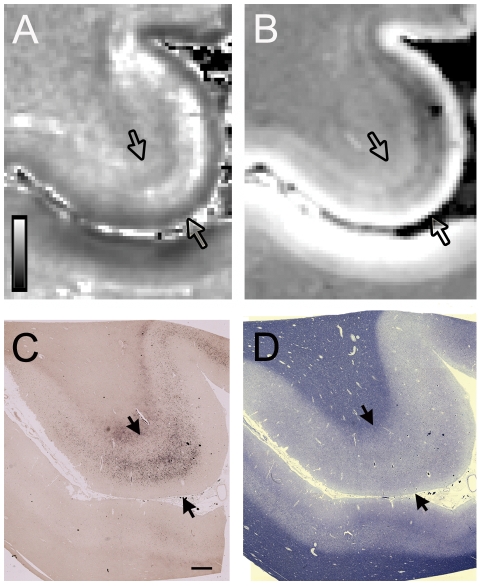
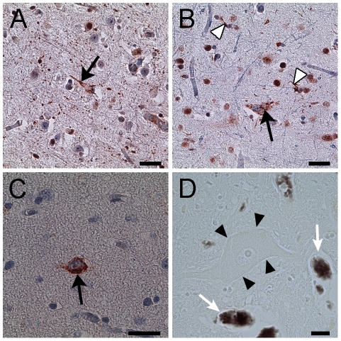
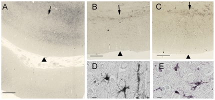
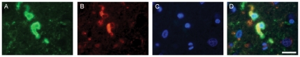
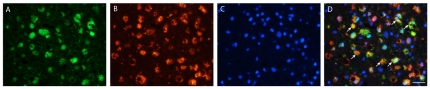
Amyotrophic lateral sclerosis (ALS) is a progressive neurodegenerative disorder characterized by cortical and spinal motor neuron dysfunction. Routine magnetic resonance imaging (MRI) studies have previously shown hypointense signal in the motor cortex on T(2)-weighted images in some ALS patients, however, the cause of this finding is unknown. To investigate the utility of this MR signal change as a marker of cortical motor neuron degeneration, signal abnormalities on 3T and 7T MR images of the brain were compared, and pathology was obtained in two ALS patients to determine the origin of the motor cortex hypointensity. Nineteen patients with clinically probable or definite ALS by El Escorial criteria and 19 healthy controls underwent 3T MRI. A 7T MRI scan was carried out on five ALS patients who had motor cortex hypointensity on the 3T FLAIR sequence and on three healthy controls. Postmortem 7T MRI of the brain was performed in one ALS patient and histological studies of the brains and spinal cords were obtained post-mortem in two patients. The motor cortex hypointensity on 3T FLAIR images was present in greater frequency in ALS patients. Increased hypointensity correlated with greater severity of upper motor neuron impairment. Analysis of 7T T(2)(*)-weighted gradient echo imaging localized the signal alteration to the deeper layers of the motor cortex in both ALS patients. Pathological studies showed increased iron accumulation in microglial cells in areas corresponding to the location of the signal changes on the 3T and 7T MRI of the motor cortex. These findings indicate that the motor cortex hypointensity on 3T MRI FLAIR images in ALS is due to increased iron accumulation by microglia.

Trial registration: ClinicalTrials.gov NCT00334516.


Conflict of interest statement

Competing Interests: The authors have declared that no competing interests exist.

Figures

Figure 1
Figure 1. 3T and 7T MRI in ALS and healthy control.
3T and 7T brain MRI signal changes in the motor cortex hand knob differed between ALS patients (A, B, C) and healthy controls (D, E, F). Cortical hypointensity (arrows) is shown to be present in an ALS patient (Patient 2, age 51) and not in a healthy control on 3T FLAIR (A, D) and 7T gradient echo magnitude images (B, E). These areas corresponded to hyperintensity in the 7T R2 * maps (C, F). Scale bar represents R2 * value in Hz.
Figure 2
Figure 2. Frequency of hypointensity grade in the hand knob of the motor cortex.
(A) Higher grades of hypointensity in the hand knob region on 3T FLAIR MRI occurred with greater frequency in ALS patients (black bars) than in healthy controls (gray bars). (B) Higher grades of hypointensity on 3T FLAIR MRI was associated with higher upper motor neuron impairment score (UMN-IS). Each dot represents the grade for one hemisphere and the UMN-IS score for the contralateral limb. Line indicates mean for group. Hypointensity was rated grade 0 (absent), grade 1 (present, mild), and grade 2 (present, marked). The UMN-IS is graded from 0–5 with normal function = 0, and higher scores indicating greater UMN impairment.
Figure 3
Figure 3. Ex vivo 7T MRI and corresponding pathology in ALS.
7T MRI signal change corresponded to iron accumulation in deeper layers of the cortical gray in the hand knob in an ALS patient. Each panel shows an axial slice of the precentral gyrus, with the cortical gray matter boundaries indicated by arrows, and the central sulcus and postcentral gyrus below. (A) R2 * map showed patchy hyperintensity in the precentral gray. Scale bar represents R2 * value in Hz. (B) Gradient echo magnitude image showed hypointensity in the corresponding region. Pixels correlate to acquisition voxels. (C) Low power micrographs show iron accumulation in the middle and deep layers of the motor cortex with Perls' DAB stain. (D) The luxol fast blue stain showed myelin pallor in the subcortical white matter of the precentral gyrus compared to the postcentral gyrus. Scale bar, 1 mm.
Figure 4
Figure 4. Immunohistochemistry of cellular inclusions and iron histology in an ALS patient.
(A) Ubiquitin reactive neuronal cytoplasmic inclusion with a filamentous appearance (arrow) in motor cortical neuron. (B, C) The neuronal cytoplasmic inclusions (arrows) were reactive with TDP-43 (B) and phosphorylated TDP-43 (C). Glial cell inclusions (B, white arrow heads) were also TDP-43 positive. (D) Spinal motor neurons (arrow heads) did not show iron accumulation with Perls' DAB stain which was present in erythrocytes in the same section (arrows). Scale bar, 100 mm (A–C); 20 mm (D).
Figure 5
Figure 5. Histological staining for iron with Perl's DAB stain in the motor cortex.
The panels show comparable planes of section through the precentral gyrus of brains from the second ALS patient (A), and from the Alzheimer (B) and Parkinson (C) patients. The arrowheads indicate the pial surface. The arrows indicate the gray-white junction. (A) Low power magnification showing iron accumulation in the middle and deeper layers of cortical gray matter, and at the gray-white junction in a 51-year-old ALS patient. At higher power (D, E) the staining is present in cells with irregular processes suggestive of microglia in the ALS motor cortex. The lower portion of panel A shows the post-central gyrus with relatively little iron staining. The iron staining was more diffuse within the motor cortex of Alzheimer (B) and Parkinson (C) patients and was also present in the subcortical white matter. Scale bars, 1 mm (A–C), 10 µm (D, E).
Figure 6
Figure 6. Double immunofluorescent labeling of ferritin-rich microglia in the motor cortex of an ALS patient.
Immunostaining for ferritin in green (A) and CD68, a marker of microglia, in red (B) labeling showed co-localization in yellow (D, merge) indicating increased microglial ferritin in the motor cortex of an ALS patient. (C) DAPI immunofluorescence, in blue, shows nuclear labeling. Scale bar, 20 mm.
Figure 7
Figure 7. Double immunofluorescent labeling of ferritin-rich microglia in the dorsolateral white matter from the spinal cord of an ALS patient.
Immunostaining for ferritin in green (A) and CD68, a marker of microglia, in red (B) labeling showed co-localization (arrows) in yellow (D, merge) indicating increased microglial ferritin in the corticospinal tract from the spinal cord of an ALS patient. (C) DAPI immunofluorescence, in blue, shows nuclear labeling. Scale bar, 30 mm.

References

    1. Ince PG, Evans J, Knopp M, Forster G, Hamdalla HH, et al. Corticospinal tract degeneration in the progressive muscular atrophy variant of ALS. Neurology. 2003;60:1252–1258. - PubMed
    1. Ellis CM, Simmons A, Jones DK, Bland J, Dawson JM, et al. Diffusion tensor MRI assesses corticospinal tract damage in ALS. Neurology. 1999;53:1051–1058. - PubMed
    1. Mitsumoto H, Ulug AM, Pullman SL, Gooch CL, Chan S, et al. Quantitative objective markers for upper and lower motor neuron dysfunction in ALS. Neurology. 2007;68:1402–1410. - PubMed
    1. Iwata NK, Kwan JY, Danielian LE, Butman JA, Tovar-Moll F, et al. White matter alterations differ in primary lateral sclerosis and amyotrophic lateral sclerosis. Brain. 2011;134:2642–2655. - PMC - PubMed
    1. Hecht MJ, Fellner F, Fellner C, Hilz MJ, Neundorfer B, et al. Hyperintense and hypointense MRI signals of the precentral gyrus and corticospinal tract in ALS: a follow-up examination including FLAIR images. J Neurol Sci. 2002;199:59–65. - PubMed

Publication types

MeSH terms

Associated data