. 2012;7(4):e35282.
doi: 10.1371/journal.pone.0035282. Epub 2012 Apr 17.

Agouti revisited: transcript quantification of the ASIP gene in bovine tissues related to protein expression and localization

Affiliations

Agouti revisited: transcript quantification of the ASIP gene in bovine tissues related to protein expression and localization

Elke Albrecht et al. PLoS One. 2012.

Abstract

Beside its role in melanogenesis, the agouti signaling protein (ASIP) has been related to obesity. The potentially crucial role in adipocyte development makes it a tempting candidate for economic relevant, fat related traits in farm animals. The objective of our study was to characterize the mRNA expression of different ASIP transcripts and of putative targets in different bovine tissues, as well as to study consequences on protein abundance and localization. ASIP mRNA abundance was determined by RT-qPCR in adipose and further tissues of cattle representing different breeds and crosses. ASIP mRNA was up-regulated more than 9-fold in intramuscular fat of Japanese Black cattle compared to Holstein (p<0.001). Further analyses revealed that a transposon-derived transcript was solely responsible for the increased ASIP mRNA abundance. This transcript was observed in single individuals of different breeds indicating a wide spread occurrence of this insertion at the ASIP locus in cattle. The protein was detected in different adipose tissues, skin, lung and liver, but not in skeletal muscle by Western blot with a bovine-specific ASIP antibody. However, the protein abundance was not related to the observed ASIP mRNA over-expression. Immuno-histochemical analyses revealed a putative nuclear localization of ASIP additionally to the expected cytosolic signal in different cell types. The expression of melanocortin receptors (MCR) 1 to 5 as potential targets for ASIP was analyzed by RT-PCR in subcutaneous fat. Only MC1R and MC4R were detected indicating a similar receptor expression like in human adipose tissue. Our results provide evidence for a widespread expression of ASIP in bovine tissues at mRNA and, for the first time, at protein level. ASIP protein is detectable in adipocytes as well as in further cells of adipose tissue. We generated a basis for a more detailed investigation of ASIP function in peripheral tissues of various mammalian species.


Conflict of interest statement

Competing Interests: The authors have declared that no competing interests exist.

Figures

Figure 1
Figure 1. ASIP mRNA abundance in M. longissimus (MLD) and dissected intramuscular fat (IMF).
Bars represent means of fold changes compared to Holstein (n = 5) with 95% confidence interval, marked by vertical lines. Different letters indicate significant differences to Holstein within tissue (p<0.05). Number of samples: Japanese Black (n = 6 [MLD], n = 5 [IMF]), Charolais (n = 6).
Figure 2
Figure 2. Structure of the bovine ASIP locus and resulting transcripts.
(A) Non-coding (gray) and coding exons of ASIP (black) are given as boxes and are numbered below. Exons in parentheses were not observed in this study. Smaller numbers indicate exon and intron sizes in base pairs. A LINE element (L1-BT) is inserted between non-coding and coding exons. The underlying sequence (GenBank accession no. GK000013.2) contains two gaps. The size of the first gap was determined by sequencing whereas the second gap was closed in silico by insertion of partial sequence from DQ000238.1. (B) Transcripts of the bovine ASIP gene resulting from different use of non-coding exons. Transcript 2C recruits a non-coding exon from the LINE. Transcript 1A was not observed in our study. The figure was modified and supplemented on the basis of data from Girardot et al. , .
Figure 3
Figure 3. ASIP mRNA abundance (all transcripts) and abundance of transcript 2C in bovine intramuscular fat (IMF).
Bars represent individual Cp from qPCR in IMF of Holstein, Japanese Black and Charolais steers normalized to 2 reference genes. Note that high ASIP mRNA abundance (low Cp) was observed in samples with expression of transcript 2C only.
Figure 4
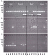
Figure 4. Expression of ASIP transcripts in different tissues of crossbred bulls.
All transcripts were amplified in cDNA (40 cycles) from the tissues indicated on panel (A). The band intensity is not indicative for mRNA abundance. (A) Transcript 1B (157 bp). A repeated amplification is shown in the framed box indicating poor reproducibility in liver cDNA. (B) Transcript 2C (244 bp) is detectable in all tissues of bull #4. Sequencing of the product obtained in liver cDNA of bull #5 failed. (C) Skin specific transcript 1C (132 bp). The larger band observed in bull #4 was identified as transcript 1C2C (293 bp) by sequencing. M: molecular weight marker.
Figure 5
Figure 5. ASIP protein expression in different tissues of bulls with normal expression (−) or over-expression of ASIP mRNA (+).
Chemiluminescence detection of ASIP and β-actin by Western blotting of 40 µg protein of the respective tissues. Lanes 1 and 2: M. longissimus, 3 and 4: subcutaneous fat, 5 and 6: intermuscular fat, 7 and 8: heart, 9 and 10: liver, 11 and 12: lung.
Figure 6
Figure 6. Cellular localization of ASIP protein in different bovine tissues.
The left panels show the overlay of Hoechst 33258 nuclear stain with the bright-field image. In the right panels, ASIP protein is labeled with Alexa 488 (green). White arrows indicate distinct signals located at nuclei. (A) M. longissimus with included intramuscular fat. (B) cardiac muscle, (C) subcutaneous fat, (D) intermuscular fat, (E) liver, (F) lung.
Figure 7
Figure 7. Expression of melanocortin receptors 1–5 (MCR) in subcutaneous fat (SCF) of crossbred bulls.
All MCR were amplified in cDNA from SCF (40 cycles). Specific PCR products were obtained for MC1R (233 bp) and MC4R (163 bp). +: PCR product obtained in genomic DNA. M: molecular weight marker.

Similar articles

Cited by

References

    1. Bultman SJ, Russell LB, Gutierrez-Espeleta GA, Woychik RP. Molecular characterization of a region of DNA associated with mutations at the agouti locus in the mouse. Proc Natl Acad Sci U S A. 1991;88:8062–8066. - PMC - PubMed
    1. Claycombe KJ, Xue BZ, Mynatt RL, Zemel MB, Moustaid-Moussa N. Regulation of leptin by agouti. Physiol Genomics. 2000;2:101–105. - PubMed
    1. Lu D, Willard D, Patel IR, Kadwell S, Overton L, et al. Agouti protein is an antagonist of the melanocyte-stimulating-hormone receptor. Nature. 1994;371:799–802. - PubMed
    1. Cone RD, Lu D, Koppula S, Vage DI, Klungland H, et al. The melanocortin receptors: agonists, antagonists, and the hormonal control of pigmentation. Recent Prog Horm Res. 1996;51:287–317. - PubMed
    1. Hiragaki T, Inoue-Murayama M, Miwa M, Fujiwara A, Mizutani M, et al. Recessive black is allelic to the yellow plumage locus in Japanese quail and associated with a frameshift deletion in the ASIP gene. Genetics. 2008;178:771–775. - PMC - PubMed

LinkOut - more resources