. 2012 Jul;18(13-14):1454-63.
doi: 10.1089/ten.TEA.2011.0550. Epub 2012 Jun 5.

Partial characterization of the Sox2+ cell population in an adult murine model of digit amputation

Affiliations

Partial characterization of the Sox2+ cell population in an adult murine model of digit amputation

Vineet Agrawal et al. Tissue Eng Part A. 2012 Jul.

Abstract

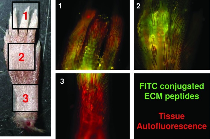
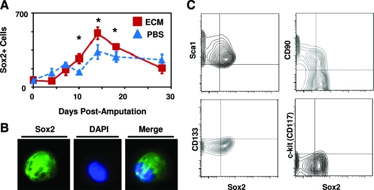
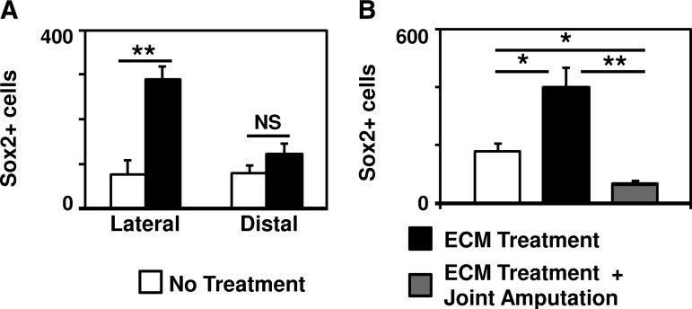
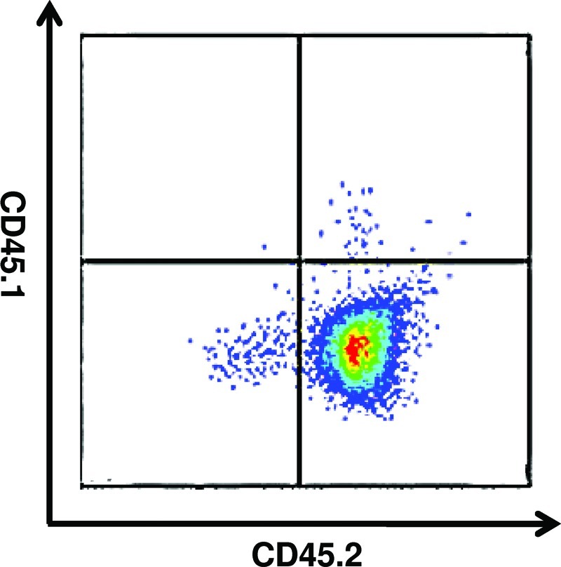
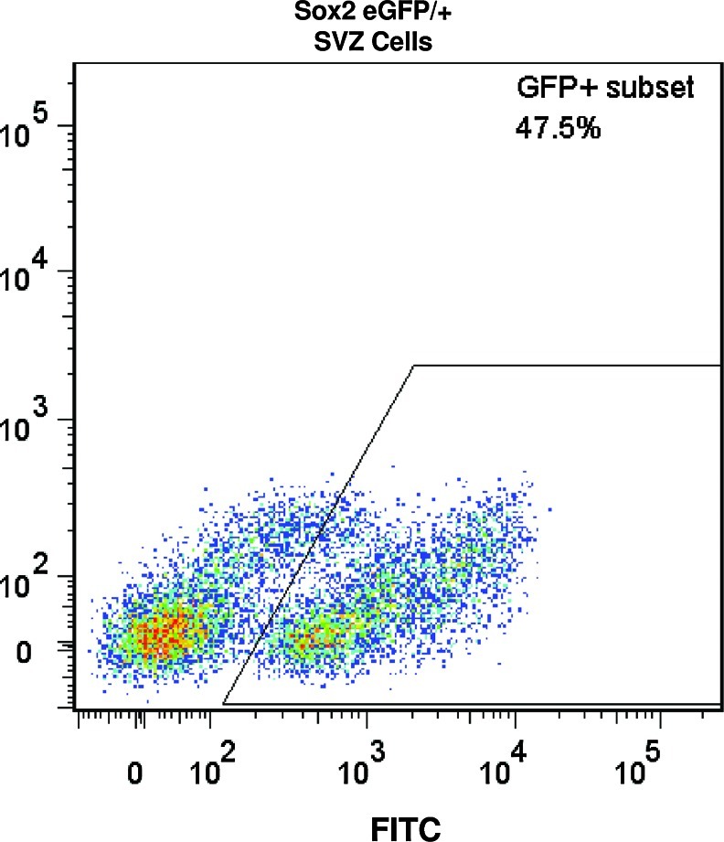
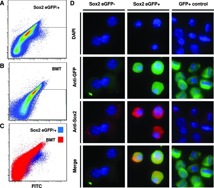
Tissue regeneration in response to injury in adult mammals is generally limited to select tissues. Nonmammalian species such as newts and axolotls undergo regeneration of complex tissues such as limbs and digits via recruitment and accumulation of local and circulating multipotent progenitors preprogrammed to recapitulate the missing tissue. Directed recruitment and activation of progenitor cells at a site of injury in adult mammals may alter the default wound-healing response from scar tissue toward regeneration. Bioactive molecules derived from proteolytic degradation of extracellular matrix (ECM) proteins have been shown to recruit a variety of progenitor cells in vitro and in vivo to the site of injury. The present study further characterized the population of cells accumulating at the site of injury after treatment with ECM degradation products in a well-established model of murine digit amputation. After a mid-second phalanx digit amputation in 6-8-week-old adult mice, treatment with ECM degradation products resulted in the accumulation of a heterogeneous population of cells, a subset of which expressed the transcription factor Sox2, a marker of pluripotent and adult progenitor cells. Sox2+ cells were localized lateral to the amputated P2 bone and coexpressed progenitor cell markers CD90 and Sca1. Transgenic Sox2 eGFP/+ and bone marrow chimeric mice showed that the bone marrow and blood circulation did not contribute to the Sox2+ cell population. The present study showed that, in addition to circulating progenitor cells, resident tissue-derived cells also populate at the site of injury after treatment with ECM degradation products. Although future work is necessary to determine the contribution of Sox2+ cells to functional tissue at the site of injury, recruitment and/or activation of local tissue-derived cells may be a viable approach to tissue engineering of more complex tissues in adult mammals.


Figures

FIG. 1.
FIG. 1.
Extracellular matrix (ECM) degradation products reach the site of amputation. After injecting fluorescein isothiocyanate-conjugated ECM degradation products at the base of the amputated digit, ECM degradation products were found diffusing along the amputated digit as well as adjacent unamputated digits. Color images available online at www.liebertpub.com/tea
FIG. 2.
FIG. 2.
ECM treatment causes a more densely cellular accumulation at the site of digit amputation. (A) After mid-second phalanx digit amputation and treatment with ECM degradation products or control, histologic analysis of Trichrome-stained sections at day 14 postamputation showed the appearance of a more densely cellular accumulation at the site of amputation after ECM treatment. (B) This was confirmed by automated quantification of the relative ratio of cellularity to connective tissue of the Trichrome stain. (C) Quantification of the area of growth distal to the site of amputation showed no difference between ECM treatment and no treatment. *p<0.05 and **p<0.01.; Error bars are mean±SD (n=4 for each group). Color images available online at www.liebertpub.com/tea
FIG. 3.
FIG. 3.
ECM treatment results in the accumulation of a heterogeneous population of cells at the site of amputation. After digit amputation and treatment with either ECM degradation products or control, the accumulation of cells at the site of amputation (Fig. 2.) was microdissected and dissociated for further analysis. Flow cytometric analysis showed that subsets of cells expressed markers Sca1, CD146, Sox2, Lineage, CD11b, and F4/80.
FIG. 4.
FIG. 4.
ECM treatment results in a greater number of Sox2+ cells at the site of amputation. (A) After digit amputation and treatment with ECM degradation products or control, digits were fixed at various time points postamputation, sectioned for histologic analysis, and immunolabeled for the presence of Sox2. A greater number of Sox2+ cells were observed at the site of amputation on days 10, 14, and 18 postamputation. (B) After microdissection and dissociation of the cell accumulation at the site of amputation, Sox2+ cell expression was confirmed by immunolabeling of cells cytospun on to a slide. (C) Flow cytometric analysis of the isolated cells showed that Sox2+ cells coexpressed Sca1 and CD90, but not CD133 or c-kit. *p<0.05. Error bars are mean±SEM (n=4 for each group). Color images available online at www.liebertpub.com/tea
FIG. 5.
FIG. 5.
Sox2+ cell accumulation requires bone injury and is located lateral to the amputated bone. (A) Immunolabeling of histologic sections of amputated digits showed that the majority of Sox2+ cells present at the site of amputation after treatment with ECM degradation products were located lateral to the amputated P2 bone, consistent with a periosteal location. (B) After digit amputation proximal to P2 bone at the joint such that no bone injury was induced, the accumulation of Sox2+ cells at the site of amputation after ECM degradation products was decreased. *p<0.05. **p<0.01. Error bars are mean±SEM (n=4 for each group).
FIG. 6.
FIG. 6.
Confirmation of stable engraftment of bone marrow transplanted mice. To confirm stable engraftment of Sox2 eGFP/+ bone marrow transplanted into adult wild type C57/BL6 mice, blood from transplanted mice was analyzed at 4 weeks postengraftment for expression of CD45.2 (Sox2 eGFP/+) and CD45.1 (wild type). At 4 weeks postengraftment, the blood of mice expressed the CD45.2 on blood-derived cells, confirming stable engraftment of Sox2/eGFP+ bone marrow in mice. Color images available online at www.liebertpub.com/tea
FIG. 7.
FIG. 7.
Confirmation of eGFP+ expression in Sox2+ cells from the subventricular zone (SVZ) of adult Sox2 eGFP/+ mice. Cells from the SVZ of the adult brain of Sox2 eGFP/+ mice were isolated to confirm eGFP+ expression. GFP+ expression was confirmed in a subset of the SVZ, as previously shown.35–37 Isolated bone marrows did not express eGFP. Color images available online at www.liebertpub.com/tea
FIG. 8.
FIG. 8.
Sox2+ cells at the site of digit amputation are not derived from the bone marrow or circulation. Sox2 eGFP/+ transgenic mice and genetically matched, wild-type C57/BL6 transplanted with Sox2 eGFP/+ bone marrow were subjected to mid-second phalanx digit amputation and treatment with ECM degradation products. At day 14 postamputation, cells at the site of amputation were microdissected and dissociated for flow cytometric analysis for GFP expression. (A) GFP+ cells were found in cells isolated from Sox2 eGFP/+ transgenic mice. (B) A GFP+ population of cells was not found in cells isolated from bone marrow chimeric wild-type mice. (C) Cells isolated from Sox2 eGFP/+ mice showed a population of cells by flow cytometric analysis that was not present in bone marrow chimeric wild-type mice. (D) After sorting and cytospinning GFP+ and GFP– cell populations, immunolabeling confirmed that the GFP+ cells expressed Sox2 and GFP, whereas GFP– cells did not express Sox2 or GFP. Color images available online at www.liebertpub.com/tea

References

    1. Kumar A. Godwin J.W. Gates P.B. Garza-Garcia A.A. Brockes J.P. Molecular basis for the nerve dependence of limb regeneration in an adult vertebrate. Science. 2007;318:772. - PMC - PubMed
    1. Monaghan J.R. Epp L.G. Putta S. Page R.B. Walker J.A. Beachy C.K., et al. Microarray and cDNA sequence analysis of transcription during nerve-dependent limb regeneration. BMC Biol. 2009;7:1. - PMC - PubMed
    1. Kragl M. Knapp D. Nacu E. Khattak S. Maden M. Epperlein H.H., et al. Cells keep a memory of their tissue origin during axolotl limb regeneration. Nature. 2009;460:60. - PubMed
    1. Kim J.Y. Xin X. Moioli E.K. Chung J. Lee C.H. Chen M., et al. Regeneration of Dental-Pulp-like Tissue by Chemotaxis-Induced Cell Homing. Tissue Eng Part A. 2010;16:3023. - PMC - PubMed
    1. Lee C.H. Cook J.L. Mendelson A. Moioli E.K. Yao H. Mao J.J. Regeneration of the articular surface of the rabbit synovial joint by cell homing: a proof of concept study. Lancet. 2010;376:440. - PMC - PubMed

Publication types

Substances