. 2012 Jul;33(21):5247-58.
doi: 10.1016/j.biomaterials.2012.03.076. Epub 2012 Apr 22.

A functional agarose-hydroxyapatite scaffold for osteochondral interface regeneration

Affiliations

A functional agarose-hydroxyapatite scaffold for osteochondral interface regeneration

Nora T Khanarian et al. Biomaterials. 2012 Jul.

Abstract

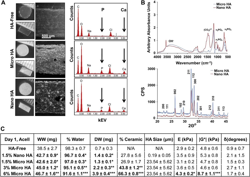
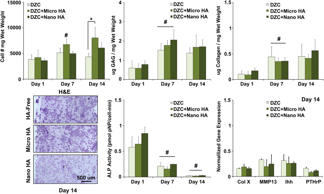
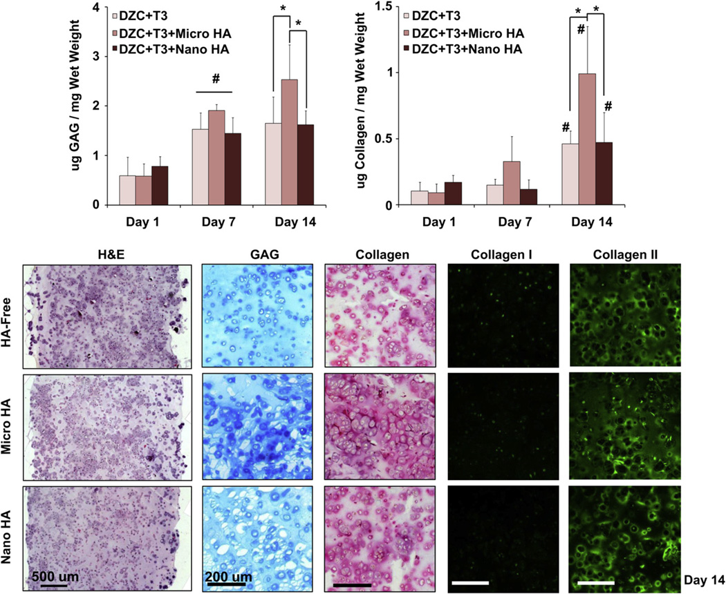
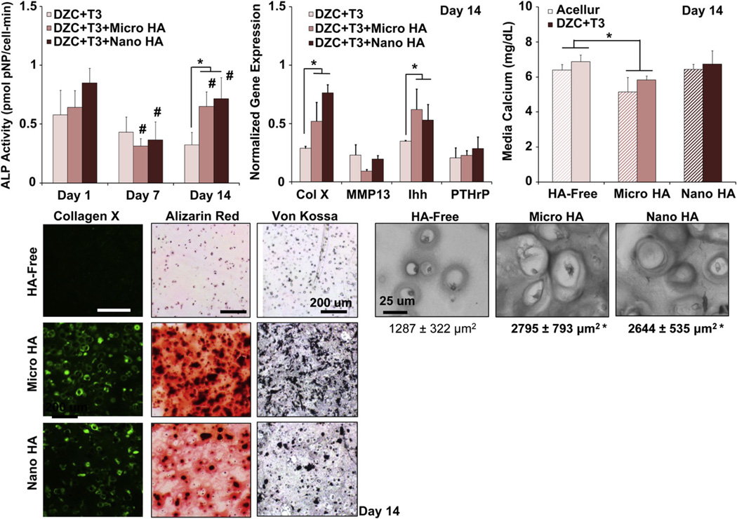
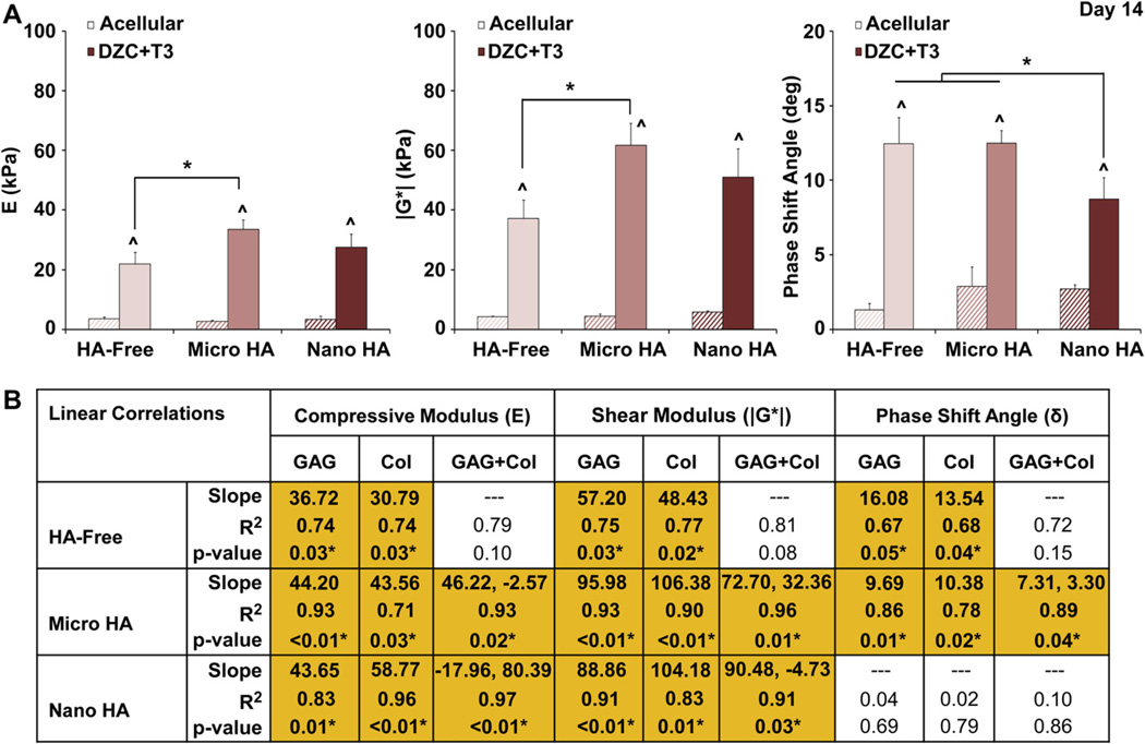
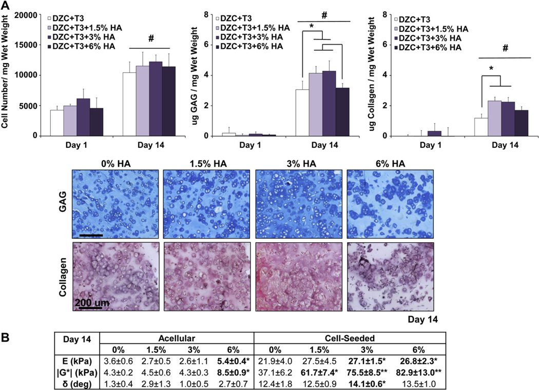
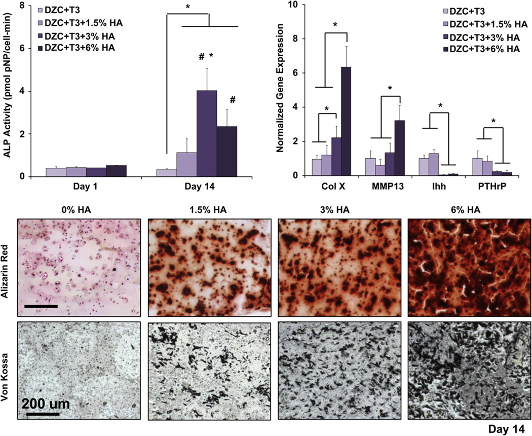
Regeneration of the osteochondral interface is critical for integrative and functional cartilage repair. This study focuses on the design and optimization of a hydrogel-ceramic composite scaffold of agarose and hydroxyapatite (HA) for calcified cartilage formation. The first study objective was to compare the effects of HA on non-hypertrophic and hypertrophic chondrocytes cultured in the composite scaffold. Specifically, cell growth, biosynthesis, hypertrophy, and scaffold mechanical properties were evaluated. Next, the ceramic phase of the scaffold was optimized in terms of particle size (200 nm vs. 25 μm) and dose (0-6 w/v%). It was observed that while deep zone chondrocyte (DZC) biosynthesis and hypertrophy remained unaffected, hypertrophic chondrocytes measured higher matrix deposition and mineralization potential with the addition of HA. Most importantly, higher matrix content translated into significant increases in both compressive and shear mechanical properties. While cell hypertrophy was independent of ceramic size, matrix deposition was higher only with the addition of micron-sized ceramic particles. In addition, the highest matrix content, mechanical properties and mineralization potential were found in scaffolds with 3% micro-HA, which approximates both the mineral aggregate size and content of the native interface. These results demonstrate that the biomimetic hydrogel-ceramic composite is optimal for calcified cartilage formation and is a promising design strategy for osteochondral interface regeneration.


Figures

Fig. 1
Fig. 1
Scaffold characterization. (A) Light microscopy images of HA-free and HA-containing scaffolds (top-view and side-view). There is no change in scaffold dimensions (n = 5) with the addition of HA particles. Environmental Scanning Electron Microscopy (ESEM, 100x) reveals uniform particle distribution and Energy Dispersive X-ray Analysis (EDAX) confirms the presence of calcium (Ca) and phosphorus (P) in the HA-containing scaffolds. (B) Fourier Infrared Spectroscopy (FTIR) and X-ray Diffraction confirms that there are no difference in chemistry and crystallinity between micro-HA and nano-HA. (C) The addition of HA resulted in a dose-dependent decrease in scaffold water content (*p < 0.05: relative to HA-free, **p < 0.05: relative to HA-free and 1.5% HA groups, ***relative to HA-free, 1.5%, and 3% HA groups). Significant increases in compressive modulus (*p < 0.05, n = 3) and magnitude of the dynamic shear modulus (***p < 0.05, n = 3) were found for the 6% HA group.
Fig. 2
Fig. 2
Effect of HA presence and size on deep zone chondrocytes. A higher cell number was measured at day 14 for the micro-HA group as compared to HA-free control (*p < 0.05, n = 5). Both cells and matrix are uniformly distributed throughout the scaffolds (H&E, 10x, bar = 500 µm, Day 14, n = 2), and GAG as well as collagen content increased for all groups over the first week of culture (#p < 0.05, n = 5). Cell ALP activity decreased over time for all groups (#p < 0.05, n = 5), with no change detected in the expression of hypertrophic markers due to either the presence or size of HA particles (n = 3).
Fig. 3
Fig. 3
Effect of HA presence and size on hypertrophic chondrocytes: biosynthesis. Both GAG and collagen content were significantly higher at day 14 for the micro-HA group (*p < 0.05, n = 5). Corresponding histology reveals strongly positive matrix staining for the micro-HA group (Alcian Blue for GAG and Picrosirius Red for collagen, 10x, bar = 200 µm, day 14, n = 2). More specifically, strongly positive collagen II staining and minimal collagen I staining, was observed for all groups (immunohistochemistry, 10x, bar = 200 µm, Day 14, n = 2).
Fig. 4
Fig. 4
Effect of HA presence and size on hypertrophic chondrocyte: mineralization. The presence of HA significantly elevated ALP activity in hypertrophic chondrocytes by day 14 (*p < 0.05, n = 5), with a corresponding increase in cell size in the HA-containing scaffolds (*p < 0.05, bar = 25 µm, 32x, n = 12). Both type X collagen and Ihh expression are upregulated by day 14 (*p < 0.05, n = 3), with positive staining for type X collagen found at day 14 in the HA-containing scaffolds independent of particle size (10x, bar = 200 µm, n = 2). A significant decrease in media calcium was only found for the acellular and cell-laden micro-HA groups as compared to plain media control (*p < 0.05, n = 5), with no difference seen due to HA size. Von Kossa and Alizarin Red staining confirm the uniform distribution of HA particles in the HA-containing scaffolds, with strongly positive mineral staining seen in the micro-HA group (10x, bar = 200 µm, Day 14, n = 2).
Fig. 5
Fig. 5
Effect of HA presence and size on hypertrophic chondrocytes: scaffold mechanical properties. (A) The micro-HA containing scaffold measured a higher compressive modulus and magnitude of the dynamic shear modulus as compared to the HA-free control (*p < 0.05, Day 14), and the addition of nano-HA resulted in a lower phase shift angle (*p < 0.05). Note that mechanical properties of all cell-laden scaffolds were significantly higher than those of the corresponding acellular controls (^p < 0.05, Day 14, n = 3). (B) For the micro-HA group, linear regression analysis revealed a strong positive correlation between scaffold mechanical properties (E, |G*|, and δ) and matrix content (GAG, Col) as well as matrix interactions (GAG + Col)(*p < 0.05, Day 14, n = 6).
Fig. 6
Fig. 6
Effect of HA dose on hypertrophic chondrocytes: growth, biosynthesis and scaffold mechanical properties. (A) While there is no dose-dependent effect on cell growth, the addition of 1.5% and 3% micro-HA resulted in higher GAG and collagen deposition by day 14 (*p < 0.05, n = 5), as confirmed by histology (Alcian Blue for GAG and Picrosirius Red for collagen 10x, bar = 200 µm, Day 14, n = 2). (B) Cell-laden scaffolds measured significantly higher compressive modulus, magnitude of the dynamic shear modulus, and phase shift angle as compared to corresponding acellular controls at day 14 (n = 3). A dose-dependent increase in scaffold compressive and shear moduli was found, with those of the 3% and 6% HA groups being significantly higher than that of the 0% HA group (*p < 0.05, n = 3). In addition, cell-laden scaffolds with 3% HA measured a higher phase shift angle as compared to the 0% HA group (*p < 0.05, n = 3).
Fig. 7
Fig. 7
Effect of HA dose on hypertrophic chondrocytes: mineralization. A dose-dependent effect in ALP activity was found, with the highest enzyme activity detected in the 3% HA groups (*p < 0.05, n = 5). Cell ALP activity also increased over time for both the 3% and 6% HA groups (#p < 0.05, n = 5). Similarly, a dose-dependent increase in type X collagen expression was found, with the highest collagen X and MMP13 expression detected in the 6% HA group by day 14. In contrast, significant suppression of PTHrP and Ihh expression was found at the higher HA doses (3% and 6% HA, *p < 0.05, n = 3 normalized to 0% group). For mineralization, both von Kossa and Alizarin Red staining revealed the presence of pre-incorporated HA particles, as well as cell-mediated mineral deposition (10x, bar = 200 µm, Day 14, n = 2).

References

    1. Hunziker EB. Biologic repair of articular cartilage. Defect models in experimental animals and matrix requirements. Clin Orthop Relat Res. 1999:S135–S146. - PubMed
    1. Frisbie DD, Trotter GW, Powers BE, Rodkey WG, Steadman JR, Howard RD, et al. Arthroscopic subchondral bone plate microfracture technique augments healing of large chondral defects in the radial carpal bone and medial femoral condyle of horses. Vet Surg. 1999;28:242–255. - PubMed
    1. Bedi A, Feeley BT, Williams RJ., III Management of articular cartilage defects of the knee. J Bone Joint Surg Am. 2010;92:994–1009. - PubMed
    1. Hunziker EB. Articular cartilage repair: basic science and clinical progress. A review of the current status and prospects. Osteoarthritis Cartilage. 2002;10:432–463. - PubMed
    1. Malmonge SM, Zavaglia CA, Belangero WD. Biomechanical and histological evaluation of hydrogel implants in articular cartilage. Braz J Med Biol Res. 2000;33:307–312. - PubMed

Publication types