

Mineralization of articular cartilage in the Sprague-Dawley rat: characterization and mechanical analysis
- PMID: 22531460
- PMCID: PMC3374052
- DOI: 10.1016/j.joca.2012.04.011
Mineralization of articular cartilage in the Sprague-Dawley rat: characterization and mechanical analysis
Abstract
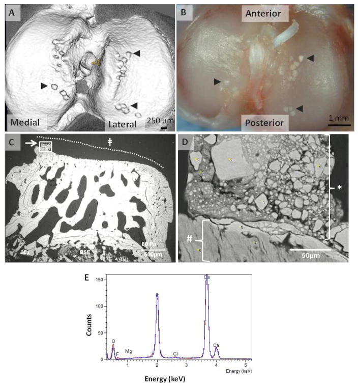
The formation of mineralized deposits in human articular cartilage is a common occurrence [–4]; however, the relationship between mineral deposition and material properties of the articular cartilage is not well understood nor the relationship between mineral deposition and the development of degenerative joint disease. Several different crystalline structures have been identified in articular cartilage and synovial fluid including monosodium urate, calcium pyrophosphate dihydrate (CPPD), and basic calcium phosphates (BCPs). These distinct mineral phases are associated with specific pathologies and mechanisms of crystal formation such as the development of monosodium urate in gout and CPPD in pseudogout. Less is known regarding the deposition of BCPs, a class of compounds including carbonate-substituted hydroxyapatite (cHA), tricalcium phosphates (TCP), octacalcium phosphate (OCP), and whitlockite, in articular cartilage. The presence of BCP calcification of articular cartilage in humans has been associated with decreased joint function [1, 3], aging [2] and severity of osteoarthritis [1, 3]. Commonly used methods of crystal detection such as polarized light microscopy of synovial fluid and conventional radiography of the joint can be insensitive to the detection of BCP crystals and more sensitive techniques such as microradiography or electron microscopy of articular cartilage sections are required to detect areas of BCP mineralization [3, 5, 6]. It is not yet known how regions of mineralization may influence the tribological properties (friction, wear, and lubrication) of the articulating surfaces and the material and structural properties of articular cartilage. Animal models with which to study the mechanisms of mineralization of articular cartilage are limited.
Conflict of interest statement
Maria Roemhildt, Ph.D. - has no competing interests
Bruce Beynnon, Ph.D. - has no competing interests
Mack Gardner-Morse, M.S. - has no competing interests
Figures
Similar articles
-
Articular chondrocalcinosis of the humeral head in greyhounds.Am J Vet Res. 1995 Apr;56(4):473-80. Am J Vet Res. 1995. PMID: 7785825
-
Effect of Vertical or Beveled Chondral Defect Creation on Rim Deformation and Contact.Cartilage. 2019 Apr;10(2):222-228. doi: 10.1177/1947603517752058. Epub 2018 Jan 17. Cartilage. 2019. PMID: 29338324 Free PMC article.
-
[The nature of intra-articular and extra-articular localization of microcrystals in chondrocalcinosis].Cesk Radiol. 1987 Oct;41(5):301-12. Cesk Radiol. 1987. PMID: 3677189 Slovak. No abstract available.
-
Articular cartilage calcification and matrix vesicles.Curr Rheumatol Rep. 2002 Jun;4(3):265-9. doi: 10.1007/s11926-002-0075-0. Curr Rheumatol Rep. 2002. PMID: 12010613 Review.
-
Parallels between arterial and cartilage calcification: what understanding artery calcification can teach us about chondrocalcinosis.Curr Opin Rheumatol. 2003 May;15(3):302-10. doi: 10.1097/00002281-200305000-00019. Curr Opin Rheumatol. 2003. PMID: 12707585 Review.
Cited by
-
Cartilage calcification of the ankle joint is associated with osteoarthritis in the general population.BMC Musculoskelet Disord. 2018 May 24;19(1):169. doi: 10.1186/s12891-018-2094-7. BMC Musculoskelet Disord. 2018. PMID: 29793463 Free PMC article.
-
Total-body irradiation produces late degenerative joint damage in rats.Int J Radiat Biol. 2014 Sep;90(9):821-30. doi: 10.3109/09553002.2014.927935. Epub 2014 Aug 11. Int J Radiat Biol. 2014. PMID: 24885745 Free PMC article.
-
The Relationship between Vitamin K and Osteoarthritis: A Review of Current Evidence.Nutrients. 2020 Apr 25;12(5):1208. doi: 10.3390/nu12051208. Nutrients. 2020. PMID: 32344816 Free PMC article. Review.
-
Ultrashort Time to Echo Magnetic Resonance Evaluation of Calcium Pyrophosphate Crystal Deposition in Human Menisci.Invest Radiol. 2019 Jun;54(6):349-355. doi: 10.1097/RLI.0000000000000547. Invest Radiol. 2019. PMID: 30688685 Free PMC article.
-
Age-related changes in the cartilage of the temporomandibular joint.Geroscience. 2020 Jun;42(3):995-1004. doi: 10.1007/s11357-020-00160-w. Epub 2020 Jan 28. Geroscience. 2020. PMID: 31993924 Free PMC article.
References
-
- Fuerst M, Bertrand J, Lammers L, Dreier R, Echtermeyer F, Nitschke Y, et al. Calcification of articular cartilage in human osteoarthritis. Arthritis & Rheumatism. 2009;60:2694–703. - PubMed
-
- Scotchford CA, Vickers M, Yousuf Ali S. The isolation and characterization of magnesium whitlockite crystals from human articular cartilage. Osteoarthritis and Cartilage. 1995;3:79–94. - PubMed
Publication types
MeSH terms
Grants and funding
LinkOut - more resources
Full Text Sources
Medical
