. 2012 Jul;44(7):1166-73.
doi: 10.1016/j.biocel.2012.04.005. Epub 2012 Apr 16.

The zinc finger transcription factor ZKSCAN3 promotes prostate cancer cell migration

Affiliations

The zinc finger transcription factor ZKSCAN3 promotes prostate cancer cell migration

Xingding Zhang et al. Int J Biochem Cell Biol. 2012 Jul.

Abstract

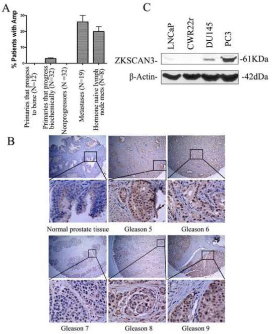
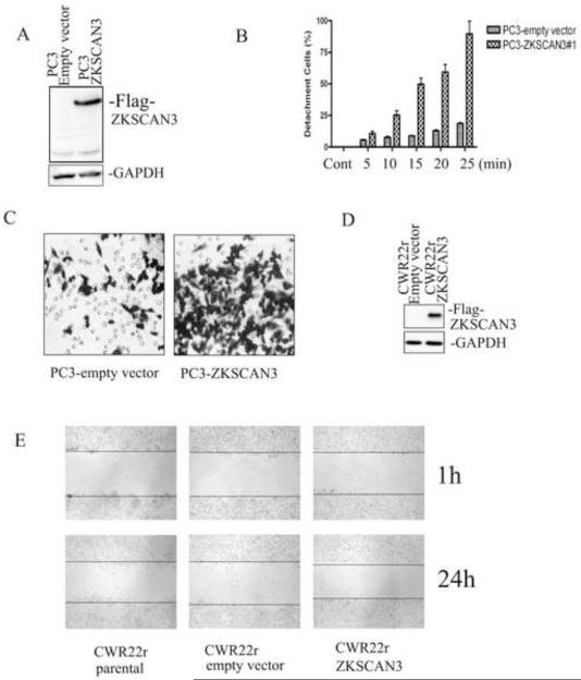
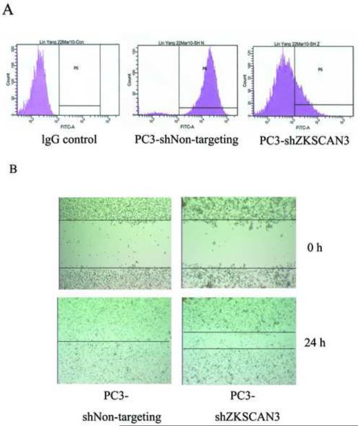
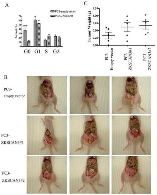
In our previous studies, ZKSCAN3 was demonstrated to be over-expressed in invasive colonic tumor cells and their liver metastases, but minimally expressed in adjacent non-transformed tissues. Further preliminary data showed that ZKSCAN3 was expressed in a majority of prostate cancer patient samples, but not in normal prostate tissues. Moreover, the ZKSCAN3 protein is highly expressed in the PC3 prostate cancer cell line, which has high metastatic potential, but little expression was observed in non-metastatic prostate cancer cell lines. Thus, we hypothesized that ZKSCAN3 could participate in tumor metastasis by regulating tumor cell migration. To test this hypothesis, ZKSCAN3 mRNA was knocked down by ZKSCAN3 specific shRNA in PC3 cells and a significant decrease in cell motility was observed. In contrast, when ZKSCAN3 cDNA was overexpressed in PC3 cells, cell detachment was observed and suspension culture induced apoptosis was greatly decreased, suggesting that ZKSCAN3 is able to enhance PC3 cell survival under anoikis stress. Additional wound healing and invasion assays showed that cell migration was enhanced by ZKSCAN3 expression. Interestingly, the ZKSCAN3 gene was amplified in 26% (5/19) of metastatic prostate cancers and 20% (1/5) of lymph node metastases, but there was no amplification found in primary prostate cancers, further supporting the role of ZKSCAN3 in tumor cell migration. In vivo studies using orthotopic tumor models indicated that overexpression of ZKSCAN3 significantly enhanced tumorigenicity. Taken together, we provide evidence that ZKSCAN3, a zinc finger transcription factor, plays a critical role in promoting prostate cancer cell migration.


Figures

Fig. 1
Fig. 1
Expression of ZKSCAN3 in primary prostate cancer, metastatic prostate cancer, and prostate cancer cell lines. A: ZKSCAN3 loci are amplified in metastatic prostate cancer. BAC arrays were used to detect ZKSCAN3 gene amplification using DNA isolated from primary or metastatic prostate cancers. The prostate cancers were divided into 5 groups: primaries that progress to bone (N=12), primaries that progress biochemically (N=32), nonprogressors (N=32), bone metastases (N=19) and hormone naïve lymph node metastases (N=8). B: Expression and localization of the ZKSCAN3 protein to the tumor cells in resected prostate cancer. Normal prostate tissue and 5 different gleasons cancer tissue were arrayed by immunohistochemistry using the affinity-purified anti-ZKSCAN3 antibody. C: The expression of ZKSCAN3 in representative prostate cancer cell lines with differential metastatic potential. ZKSCAN3 expression was analyzed in nuclear extracts from LNCaP, CWR22r, DU145 and PC3 cells by Western blotting with the anti-ZKSCAN3 antibody.
Fig. 2
Fig. 2
Exogenous expression of ZKSCAN3 increases prostate cancer cell detachment and motility. A: Generation of stable PC3-ZKSCAN3 cell lines. PC3 cells were transfected with the Flag-tagged ZKSCAN3 expression construct. Cells were selected with 0.5 mg/ml G418 and after 2 weeks, a G418-resistant pool was harvested and analyzed for ZKSCAN3 expression by Western blotting using the anti-Flag M2 antibody. B: ZKSCAN3 enhanced PC3 cell detachment. PC3-vector and PC3-ZKSCAN3 stable cells were seeded in 24 well plates. After 48 hours, the cells were treated with 2mM EDTA for 5 to 25 minutes. The cells were washed with PBS and then incubated with MTT reagent diluted in RPMI medium (5 mg/ml) at 37°C for 2 hours. The formazan crystals produced by metabolism of MTT were reconstituted in DMSO, and the optical density at 570 nm was determined using an ELISA reader. The percentage of detached cells was calculated using the formula: 100 - [(OD570 of EDTA treated cells) / (OD570 of untreated cells) × 100%]. C: ZKSCAN3 enhanced PC3 cell motility in a transwell migration assay. PC3-vector and PC3-ZKSCAN3 stable cells were added to the upper chamber of each well and allowed to migrate through the collagen coated membrane for 2 to 3 hours. Cell motility was quantified by counting the cells that migrated to the lower surface of the membrane per square milliliter using bright-field optics. D: Overexpression of Flag-tagged ZKSCAN3 was confirmed in CWR22r cells. CWR22r cells were transfected with the Flag-tagged ZKSCAN3 expression construct, and ZKSCAN3 expression was analyzed by Western blotting using the anti-Flag M2 antibody. E: Overexpression of ZKSCAN3 enhanced CW22Rr cell migration in a wound healing assay. CWR22r cells were transfected with empty-vector or ZKSCAN3 for 48 hours. A 1 mm thick wound was created using a 10 μL micropipette tip, and cell migration was photographed after 24 hours.
Fig. 3
Fig. 3
Knockdown of ZKSCAN3 decreased PC3 cell migration A: The expression of ZKSCAN3 in PC3 cells was greatly reduced by shZKSCAN3. Flow cytometry analysis was used to detect ZKSCAN3 expression in PC3 cells treated with shNon-targeting and shZKSCAN3. Permeabilized cells were stained with either an anti-ZKSCAN3 antibody or an isotype matched control antibody, followed by anti-mouse IgG1 conjugated to FITC. The expression of ZKSCAN3 was greatly inhibited by shZKSCAN3 compared to the shNon-targeting control. B: Knockdown of ZKSCAN3 enhanced PC3 cell migration in a wound healing assay. PC3 cells were transfected with shNon-targeting control or shZKSCAN3 for 48 hours. A 1 mm thick wound was created using a 10 μL micropipette tip, and cell migration was photographed after 24 hours.
Fig. 4
Fig. 4
Effect of ZKSCAN3 on cell cycle and orthotopic tumor growth. A: Cell cycle profiles were obtained in PC3 cells transfected with either ZKSCAN3 or the control empty vector by staining with propidium iodide followed by flow cytometry analysis. A total of 20,000 events were collected from each of three replicate experiments, and cell cycle distributions were analyzed using Cell Fit software (Becton Dickinson). Statistical comparisons of the differences in the cell cycle distributions were carried out by ANOVA followed by Dunnett’s t-test. **p< 0.01 PC3-ZKSCAN3 group vs. PC3-empty vector group. B: Effect of ZKSCAN3 overexpression on orthotopic tumor growth. PC3 empty vector or PC3 ZKSCAN3 transfected cells (1×10⁵ cells/25μl) were injected into the prostate of each nude mouse. After 4 weeks mice were sacrificed, and tumors were photographed. The tumors are circumscribed in the photographs. C: Orthotopic tumors were surgically removed and weighed.

References

    1. Chambers A. The metastatic process: basic research and clinical implications. Oncol Res. 1999;11:161–168. - PubMed
    1. Grimstad I. Direct evidence that cancer cell locomotion contributes importantly to invasion. Exp Cell Res. 1987;173:515–523. - PubMed
    1. Hendrix M. Editorial: potential clinical use of exploiting metastasis suppressor genes to regulate prostatic cancer. J Urol. 2003;169:1134. - PubMed
    1. Howlader N, Noone A, Krapcho M, Neyman N, Aminou R, Waldron W, et al. SEER Cancer Statistics Review, 1975-2008. National Cancer Institute; Bethesda: 2011.
    1. Janer M, Friedrichsen D, Stanford J, Badzioch M, Kolb S, Deutsch K, et al. Genomic scan of 254 hereditary prostate cancer families. Prostate. 2003;57:309–319. - PubMed

Publication types

Substances