. 2012 Mar;318(2):109-22.
doi: 10.1002/jezb.21449.

Unilateral and bilateral expression of a quantitative trait: asymmetry and symmetry in coronal craniosynostosis

Affiliations

Unilateral and bilateral expression of a quantitative trait: asymmetry and symmetry in coronal craniosynostosis

Yann Heuzé et al. J Exp Zool B Mol Dev Evol. 2012 Mar.

Abstract

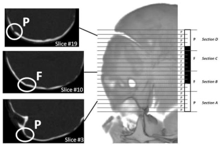
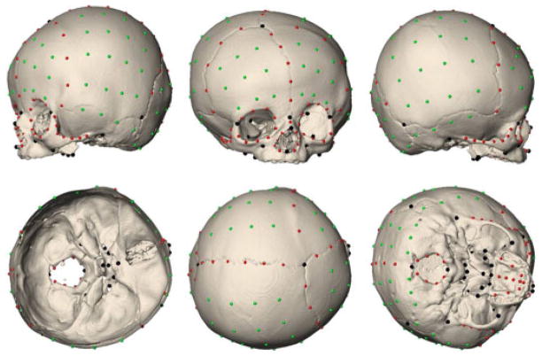
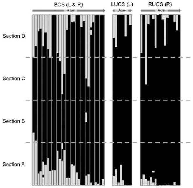
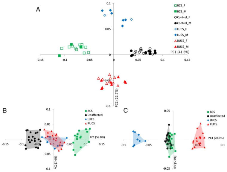
Bilateral symmetry in vertebrates is imperfect and mild asymmetries are found in normal growth and development. However, abnormal development is often characterized by strong asymmetries. Coronal craniosynostosis, defined here as consisting of premature suture closure and a characteristic skull shape, is a complex trait. The premature fusion of the coronal suture can occur unilaterally associated with skull asymmetry (anterior plagiocephaly) or bilaterally associated with a symmetric but brachycephalic skull. We investigated the relationship between coronal craniosynostosis and skull bilateral symmetry. Three-dimensional landmark coordinates were recorded on preoperative computed tomography images of children diagnosed with coronal nonsyndromic craniosynostosis (N = 40) and that of unaffected individuals (N = 20) and analyzed by geometric morphometrics. Our results showed that the fusion pattern of the coronal suture is similar across individuals and types of coronal craniosynostosis. Shape analysis showed that skulls of bilateral coronal craniosynostosis (BCS) and unaffected individuals display low degrees of asymmetry, whereas right and left unilateral coronal craniosynostosis (UCS) skulls are asymmetric and mirror images of one another. When premature fusion of the coronal suture (without taking into account cranial dysmorphology) is scored as a qualitative trait, the expected relationship between trait frequency and trait unilateral expression (i.e. negative correlation) is confirmed. Overall, we interpret our results as evidence that the same biological processes operate on the two sides in BCS skulls and on the affected side in UCS skulls, and that coronal craniosynostosis is a quantitative trait exhibiting a phenotypic continuum with BCS displaying more intense shape changes than UCS.


Figures

Figure 1
Figure 1
Diagram illustrating the method used to code coronal suture fusion. In this example, the left side of the coronal suture corresponds to 20 CT slices. The coronal suture visualized on each CT slice is scored as patent (P, white) or fused (F, black) according to the coronal suture condition. The 20 slices are then divided in four equal sections of five slices each and the sections are coded as patent or fused according to the mode of the distribution of the scores of their slices.
Figure 2
Figure 2
Illustration of the 209 points measured on 3D reconstruction of the CT images of each individual in our sample (LUCS skull shown here). Anatomical landmarks are shown in black, curve semilandmarks are shown in red, and surface semilandmarks are shown in green.
Figure 3
Figure 3
Coronal suture fusion patterns by phenotype and age. Each bar represents one side of the coronal suture. For the BCS cases, each doublet of bars represents the left, then right suture (the two bars corresponding to one individual being framed in grey). Only the affected side is shown for the UCS cases. The white and black sections represent the slices where the coronal suture was patent and fused, respectively. Though coronal suture can be represented by 20 or 100 slices depending on the CT slice thickness, all bars have been resized to a common height.
Figure 4
Figure 4
(A) Placement of the individuals on PC1 and PC2 in the shape space (principal components analysis of the Procrustes shape coordinates using all landmarks and semilandmarks of the 60 individuals). Boys are denoted by filled symbols while empty symbols denote girls. Unaffected, LUCS, RUCS, and BCS skulls are represented by circles, diamonds, triangles, and squares, respectively. (B) Placement of the individuals on PC1 and PC2 when the symmetric component of shape variation is studied. (C) Placement of the individuals on PC1 and PC2 when the asymmetric component of shape variation is studied.
Figure 5
Figure 5
Regression of skull size (lnCS) on age (months). Unaffected: open black circles; BCS: green open squares; LUCS: full blue diamonds; RUCS: full red triangles. Logarithmic fitting is represented for unaffected (black curve), BCS (green curve), LUCS (blue curve), and RUCS (red curve).
Figure 6
Figure 6
Procrustes average shape (PAS) of unaffected skulls (first row, black), PAS of RUCS skulls (second row, red), PAS of LUCS skulls (third row, blue), and PAS of BCS skulls (fourth row, green). A: lateral (left) view; B: anterior view; C: lateral (right) view; D: posterior view; E: superior view; F: endocranial base view; G: ectocranial base (inferior) view.
Figure 7
Figure 7
Placement of the half-skulls on PC1 and PC2 in the shape space after adjusting for allometry. Different convex hulls are drawn for the left and right bicoronal, left unicoronal, right unicoronal, and unaffected half-skull. Wireframe graphs representing the shape changes associated with positive and negative values of PC1 in lateral view and the shape changes associated with positive and negative values of PC2 in superior view are represented for each PC and superimposed in the top left corner and the bottom right corner, respectively.
Figure 8
Figure 8
Distribution of half-skull phenotypes according to PC1 scores obtained with the PCA of the half-skulls once allometry has been removed as in Figure 7.

References

    1. Albright AL, Byrd RP. Suture pathology in craniosynostosis. J Neurosurg. 1981;54:384–387. - PubMed
    1. Aldridge K, Hill CA, Austin JR, Percival C, Martinez-Abadias N, Neuberger T, Wang Y, Jabs EW, Richtsmeier JT. Brain phenotypes in two FGFR2 mouse models for Apert syndrome. Dev Dyn. 2010;239:987–997. - PMC - PubMed
    1. Arnaud E, Meneses P, Lajeunie E, Thorne JA, Marchac D, Renier D. Postoperative mental and morphological outcome for nonsyndromic brachycephaly. Plast Reconstr Surg. 2002;110:6–12. - PubMed
    1. Bookstein FL. Morphometric tools for landmark data: geometry and biology. Cambridge: Cambridge University Press; 1991.
    1. Bookstein FL. Landmark methods for forms without landmarks: morphometrics of group differences in outline shape. Med Image Anal. 1997;1:225–243. - PubMed

Publication types