. 2012 Jun 1;287(23):19736-49.
doi: 10.1074/jbc.M112.352930. Epub 2012 Apr 24.

Androgen receptor splice variants activate androgen receptor target genes and support aberrant prostate cancer cell growth independent of canonical androgen receptor nuclear localization signal

Affiliations

Androgen receptor splice variants activate androgen receptor target genes and support aberrant prostate cancer cell growth independent of canonical androgen receptor nuclear localization signal

Siu Chiu Chan et al. J Biol Chem. .

Abstract

Synthesis of truncated androgen receptor (AR) splice variants has emerged as an important mechanism of prostate cancer (PCa) resistance to AR-targeted therapy and progression to a lethal castration-resistant phenotype. However, the precise role of these factors at this stage of the disease is not clear due to loss of multiple COOH-terminal AR protein domains, including the canonical nuclear localization signal (NLS) in the AR hinge region. Despite loss of this NLS, we show that diverse truncated AR variant species have a basal level of nuclear localization sufficient for ligand-independent transcriptional activity. Whereas full-length AR requires Hsp90 and importin-β for active nuclear translocation, basal nuclear localization of truncated AR variants is independent of these classical signals. For a subset of truncated AR variants, this basal level of nuclear import can be augmented by unique COOH-terminal sequences that reconstitute classical AR NLS activity. However, this property is separable from ligand-independent transcriptional activity. Therefore, the AR splice variant core consisting of the AR NH(2)-terminal domain and DNA binding domain is sufficient for nuclear localization and androgen-independent transcriptional activation of endogenous AR target genes. Indeed, we show that truncated AR variants with nuclear as well as nuclear/cytoplasmic localization patterns can drive androgen-independent growth of PCa cells. Together, our data demonstrate that diverse truncated AR species with varying efficiencies of nuclear localization can contribute to castration-resistant PCa pathology by driving persistent ligand-independent AR transcriptional activity.


Figures

FIGURE 1.
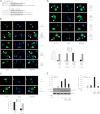
FIGURE 1.
Constitutive nuclear localization of truncated AR variants with diverse COOH-terminal extensions. A, shown is a schematic of COOH-terminal tails of truncated AR variants aligned with the AR hinge region. The multiple names that have been assigned to several of these variants are indicated (7, 8). B, Cos-7 cells expressing truncated AR variants 1/2/3/CE1, 1/2/3/CE2, 1/2/3/CE3, 1/2/3/2b, or v567es were stained with an antibody specific for the AR NTD (FITC signal), and nuclei were stained with DAPI. Stained cells were visualized by confocal microscopy. Representative images are shown for three color channels (FITC, DAPI, and merged FITC/DAPI). C, LNCaP cells expressing truncated AR variants were cultured in serum-free medium containing 1 nm mibolerone (Mib, synthetic androgen) or ethanol (EtOH, vehicle control). Cell lysates were separated into nuclear (N) and cytoplasmic (C) fractions and analyzed by Western blot with antibodies specific for the AR NTD, lamin A/C (nuclear marker), and tubulin (cytoplasmic marker). D, 22Rv1 cells were electroporated with non-targeted siRNA (CTRL) or siRNA targeted to AR Exon 7. Cells were cultured, fractionated, and analyzed by Western blot exactly as described in C.
FIGURE 2.
FIGURE 2.
Constitutive transcriptional activity of truncated AR variants with diverse COOH-terminal extensions. A, DU145 cells were transiently transfected with 4XARE-E4-LUC or MMTV-LUC reporters and expression plasmids encoding full-length AR or truncated variants 1/2/3/CE1, 1/2/3/CE2, 1/2/3/CE3, 1/2/3/2b, and v567es. Cells were cultured in serum-free medium containing 1 nm mibolerone (Mib) or vehicle control (ethanol). Cell lysates were analyzed by luciferase assay and Western blot with antibodies specific for the AR NTD and ERK-2 (loading control). Bars represent mean ± S.E. from at least three independent experiments, each performed in duplicate. Reporter activity in the absence of transactivator or ligand was arbitrarily set to 1. B, LNCaP cells were transiently transfected with a −5746 PSA-LUC reporter and truncated AR variants and analyzed by luciferase assay and Western blot as in A. C, LNCaP cells were transiently transfected and analyzed exactly as described in B with the exception of the use of AR exon 7-targeted siRNA and siRNA-resistant AR expression constructs (denoted sr (17)).
FIGURE 3.
FIGURE 3.
The COOH-terminal tail encoded by AR exon CE3 reconstitutes the canonical AR NLS. A, shown is a schematic of alanine substitutions in full-length AR and the truncated AR 1/2/3/CE3 variant. B, Cos-7 cells expressing parental and mutant plasmids illustrated in A were maintained in serum-free medium containing 1 nm mibolerone (Mib, synthetic androgen) or ethanol (vehicle control) before staining with an antibody specific for the AR NTD (FITC signal). Nuclei were stained with DAPI. Stained cells were visualized by confocal microscopy, and representative images are shown for three color channels (FITC, DAPI, and merged FITC/DAPI). Increased cytoplasmic localization is denoted by white arrows. C, Cos-7 cells were transfected with an AR 1/2/3/CE3 expression plasmid and dominant negative Ran Q69L. Cells were stained and subjected to confocal microscopy as in A. Increased cytoplasmic localization is denoted by white arrows. Quantification of predominantly nuclear (N > C) versus equal nuclear and cytoplasmic (n = C) expression is shown at the bottom. Bars represent the mean ± S.D. of 100 cells scored in three independent experiments. D, Cos-7 cells were transfected with mutant plasmids indicated in A along with dominant-negative RanQ69L (or vector control). Cells were stained and subjected to confocal microscopy as in A. Increased cytoplasmic localization is denoted by white arrows. Quantification of predominantly nuclear (N > C), equal nuclear and cytoplasmic (n = C) or predominantly cytoplasmic (N < C) expression is shown at the bottom. Bars represent mean ± S.D. of 100 cells scored in three independent experiments. E, LNCaP cells were transiently transfected with MMTV-LUC or −5746-LUC reporters and parental and mutant plasmid constructs depicted in A. Cell lysates were analyzed by luciferase assay and Western blot with antibodies specific for the AR NTD and ERK-2 (loading control). Bars represent the mean ± S.E. from at least three independent experiments, each performed in triplicate. Reporter activity in the absence of transactivator or ligand was arbitrarily set to 1.
FIGURE 4.
FIGURE 4.
NLS reconstitution does not enhance transcriptional activity of the truncated AR 1/2/3/CE2 variant. A, shown is alignment of full-length AR, AR 1/2/3/CE3, and AR 1/2/3/CE2 and location of the G629K mutation. B, left, Cos-7 cells expressing 1/2/3/CE2 and the G629K mutant were maintained in serum-free medium and stained with an antibody specific for the AR NTD (FITC signal). Nuclei were stained with DAPI. Stained cells were visualized by confocal microscopy, and representative images are shown for three color channels (FITC, DAPI, and merged FITC/DAPI). Right, shown is quantification of predominantly nuclear (N > C), equally distributed nuclear and cytoplasmic (n = C), or predominantly cytoplasmic (N < C) expression. Bars represent mean ± S.D. of 100 cells scored in 3 independent experiments. C, left, Cos-7 cells were transfected as in B with the addition of dominant-negative RanQ69L (or vector control). Right, shown is quantification of predominantly nuclear (N > C), equally distributed nuclear and cytoplasmic (n = C), or predominantly cytoplasmic (N < C) expression. Bars represent mean ± S.D. of 100 cells scored in 3 independent experiments. D, left, DU145 cells were transfected with a 4XARE-E4-LUC reporter and parental/mutant plasmid constructs depicted in A. Bars represent mean ± S.E. from at least three independent experiments, each performed in triplicate. Reporter activity in the absence of transactivator or ligand was arbitrarily set to 1. Right, cell lysates were analyzed by luciferase assay and Western blot with antibodies specific for the AR NTD and ERK-2 (loading control).
FIGURE 5.
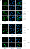
FIGURE 5.
Truncated AR variants do not require Hsp90 activity for nuclear localization. Cos-7 cells expressing full-length AR (A), AR 1/2/3/CE3 (B), or AR v567es (C) were maintained in serum-free medium containing the indicated combinations of 1 nm mibolerone (Mib, synthetic androgen), ethanol (EtOH, vehicle control), 0.8 μm 17-N-allylamino-17-demethoxygeldanamycin (17-AAG, Hsp90 inhibitor), and dimetylsulfoxide (DMSO, vehicle control) before staining with an antibody specific for the AR NTD (FITC signal). Nuclei were stained with DAPI. Stained cells were visualized by confocal microscopy, and representative images are shown for three color channels (FITC, DAPI, and merged FITC/DAPI). White arrowheads are shown to highlight cytoplasmic expression caused by 17-N-allylamino-17-demethoxygeldanamycin.
FIGURE 6.
FIGURE 6.
A unique requirement for the Arg-617/Lys-618 motif in the AR DBD terminal helix. A, shown is a schematic of the AR hinge region and location of the Arg-617/Lys-618 motif. B, DU145 cells were transiently transfected with 4XARE-E4-LUC and full-length AR or a R617A/K618A mutant. Cells were cultured in serum-free medium containing 1 nm mibolerone (Mib) or vehicle control (ethanol). Cell lysates were analyzed by luciferase assay and Western blot with antibodies specific for the AR NTD and ERK-2 (loading control). Bars represent the mean ± S.E. from at least three independent experiments, each performed in triplicate. Reporter activity in the absence of transactivator or ligand was arbitrarily set to 1. p values were derived using t tests. C, Cos-7 cells expressing parental or mutant plasmids illustrated in A were maintained in serum-free medium containing 1 nm mibolerone (Mib, synthetic androgen) or ethanol (vehicle control) before staining with an antibody specific for the AR NTD (FITC signal). Nuclei were stained with DAPI. Stained cells were visualized by confocal microscopy, and representative images are shown for three color channels (FITC, DAPI, and merged FITC/DAPI). Cytoplasmic localization resulting from the R617A/K618A mutation is denoted by white arrows. D, DU145 cells were transiently transfected with 4XARE-E4-LUC and parental/mutant truncated AR variants exactly as in B. E, Cos-7 cells were transfected with parental/mutant truncated AR variants, stained, and imaged exactly as in C. Increased cytoplasmic localization resulting from the CE3 R617A/K618A mutation is denoted by white arrows.
FIGURE 7.
FIGURE 7.
The AR NTD/DBD core is sufficient for nuclear localization and transcriptional activity of truncated AR variants. A, shown is a schematic of COOH-terminal deletions and alanine substitution mutations relative to key structural motifs in the second zinc finger of the AR DBD and hinge region. Zinc finger and α-helix locations were adapted from a prior depiction of the AR DBD crystal structure (46). B, Cos-7 cells expressing constructs depicted in A were maintained in serum-free medium and stained with an antibody specific for the AR NTD (FITC signal). Nuclei were stained with DAPI. Stained cells were visualized by confocal microscopy, and representative images are shown for three color channels (FITC, DAPI, and merged FITC/DAPI). C, DU145 cells were transiently transfected with 4XARE-E4-LUC and constructs depicted in A. Cell lysates were analyzed by luciferase assay and Western blot with antibodies specific for the AR NTD and ERK-2 (loading control). Bars represent mean ± S.E. from at least three independent experiments, each performed in triplicate. D, LNCaP cells were transiently transfected with MMTV-LUC and constructs depicted in A. Luciferase activity and transgene expression were assessed exactly as described in C. Mib, mibolerone.
FIGURE 8.
FIGURE 8.
The AR NTD/DBD core is sufficient for transcriptional activation of endogenous AR target genes. A, LNCaP cells were transduced with lentivirus encoding GFP, parental and mutant versions of AR 1/2/3/CE3, or AR 1–627. Transduced cells were maintained under serum-free conditions and stimulated with 1 nm dihydrotestosterone (DHT) or vehicle control (ethanol) for 24 h before Western blot analysis with antibodies specific for the AR NTD or ERK-2 (loading control). B, LNCaP cells were transduced exactly as in A, and RNA was subjected to quantitative RT-PCR using primer sets specific for GAPDH, PSA, hK2, TMPRSS2, and FKBP51. Expression is shown relative to GAPDH as determined using the formula 2−ΔΔCt. Bars represent the mean ± S.D. from a triplicate experiment representative of three biological replicates.
FIGURE 9.
FIGURE 9.
Truncated AR variants support biphasic, androgen-independent growth of PCa cells. A, LNCaP cells transduced with lentivirus encoding AR 1/2/3/2b, AR 1/2/3/CE3, or AR v567es were maintained in medium containing 10% charcoal-stripped (steroid-depleted) serum (10% charcoal-stripped serum). Inset, transduced cell lysates were analyzed by Western blot with antibodies specific for the AR NTD or ERK-2 (loading control). Graph, transduced cells were seeded on 24-well plates in medium containing 10% charcoal-stripped serum. At the indicated time points, cells were fixed and stained with crystal violet. Intensity of crystal violet staining (A540) was used as a surrogate of cell number. Data represent the mean ± S.D. from a quadruplicate experiment representative of three biological replicates. B, LNCaP cells were transduced with increasing doses of lentivirus encoding AR 1/2/3/CE3 and subjected to Western blot (inset) and growth assay (graph) exactly as in A. C, LNCaP cells transduced as in B were subjected to quantitative RT-PCR using primer sets specific for GAPDH, PSA, and TMPRSS2. Expression is shown relative to GAPDH as determined using the formula 2−ΔΔCt. Bars represent the mean ± S.D. from a triplicate experiment representative of three biological replicates. D, 22Rv1 cells were transduced with lentivirus encoding GFP or siRNA-resistant forms (denoted sr (17)) of AR 1/2/3/CE3, AR 1/2/3/CE3 R617A/K618A, or AR 1/2/3/CE3 K629A/R631A and electroporated with siRNA targeted to AR exon 1 as indicated. Transduced/electroporated cells were subjected to Western blot with antibodies specific for the AR NTD or ERK-2 (loading control). E, 22Rv1 cells transduced/electroporated as in D were seeded on 24-well plates in medium containing 10% charcoal-stripped serum. At the indicated time points, cells were fixed and stained with crystal violet. Intensity of crystal violet staining (A540) was used as a surrogate of cell number. Data represent the mean ± S.D. from a quadruplicate experiment representative of two biological replicates.

References

    1. Dehm S. M., Tindall D. J. (2007) Androgen receptor structural and functional elements. Role and regulation in prostate cancer. Mol. Endocrinol. 21, 2855–2863 - PubMed
    1. Knudsen K. E., Scher H. I. (2009) Starving the addiction. New opportunities for durable suppression of AR signaling in prostate cancer. Clin. Cancer Res. 15, 4792–4798 - PMC - PubMed
    1. Garraway L. A., Sellers W. R. (2006) Lineage dependency and lineage-survival oncogenes in human cancer. Nat. Rev. Cancer 6, 593–602 - PubMed
    1. de Bono J. S., Logothetis C. J., Molina A., Fizazi K., North S., Chu L., Chi K. N., Jones R. J., Goodman O. B., Jr., Saad F., Staffurth J. N., Mainwaring P., Harland S., Flaig T. W., Hutson T. E., Cheng T., Patterson H., Hainsworth J. D., Ryan C. J., Sternberg C. N., Ellard S. L., Fléchon A., Saleh M., Scholz M., Efstathiou E., Zivi A., Bianchini D., Loriot Y., Chieffo N., Kheoh T., Haqq C. M., Scher H. I., and COU-AA-301 Investigators (2011) Abiraterone and increased survival in metastatic prostate cancer. N. Engl. J. Med. 364, 1995–2005 - PMC - PubMed
    1. Dehm S. M., Tindall D. J. (2011) Alternatively spliced androgen receptor variants. Endocr. Relat. Cancer 18, R183–R196 - PMC - PubMed

Publication types

MeSH terms