

doi: 10.1093/nar/gks329.
Epub 2012 Apr 24.
Solution-state structure of an intramolecular G-quadruplex with propeller, diagonal and edgewise loops
Affiliations
- PMID: 22532609
- PMCID: PMC3413137
- DOI: 10.1093/nar/gks329
Item in Clipboard
Solution-state structure of an intramolecular G-quadruplex with propeller, diagonal and edgewise loops
Nucleic Acids Res.
2012 Aug.
Abstract
We herein report on the formation and high-resolution NMR solution-state structure determination of a G-quadruplex adopted by d[G(3)ATG(3)ACACAG(4)ACG(3)] comprised of four G-tracts with the third one consisting of four guanines that are intervened with non-G streches of different lengths. A single intramolecular antiparallel (3+1) G-quadruplex exhibits three stacked G-quartets connected with propeller, diagonal and edgewise loops of different lengths. The propeller and edgewise loops are well structured, whereas the longer diagonal loop is more flexible. To the best of our knowledge, this is the first high-resolution G-quadruplex structure where all of the three main loop types are present.
Figures
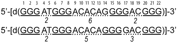
The G-rich sequence (designated ODN) investigated in this study can form two different G-quadruplexes that differ in the length of the loops. Guanines that potentially form G-quadruplex core are underlined with full line. Loop-forming residues are underlined with dotted line and the length of the loop is indicated under the loop. The nucleotides in the ODN are numbered.
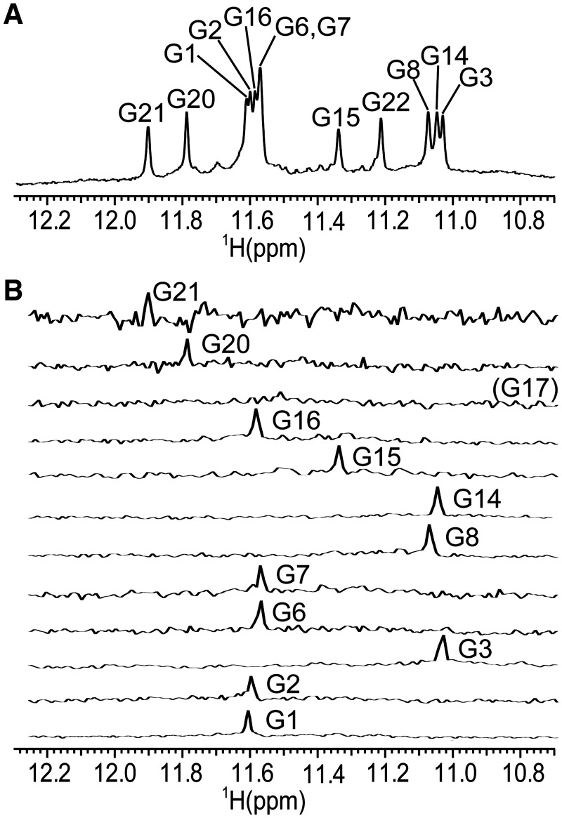
Assignment of imino proton resonances. (A) The imino region of 1D 1H-NMR spectrum and assignment of guanine residues in G-quartets. (B) 1D 15N filtered HSQC spectra of samples containing partially (6%) 15N residue-specifically labelled oligonucleotides. Spectra were recorded at 800 MHz (1D 1H-NMR spectrum) and 600 MHz (1D 15N filtered HSQC spectra) at 25°C in 10% 2H2O, 50 mM KCl, 10 mM phosphate buffer with pH 6.8. Oligonucleotide concentrations were between 1.5 and 2.7 mM.
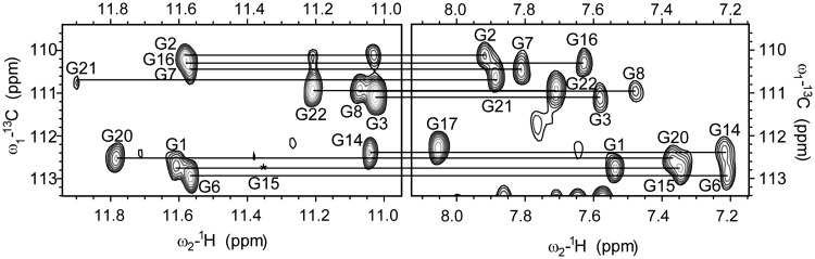
The imino and aromatic regions of 2D JRHMBC spectrum showing correlations between H8 and the corresponding imino proton with C5 of guanine residues. Missing cross-peak of G15 in the imino region is designated with asterisk. 2D JRHMBC spectrum was recorded at 800 MHz, 25°C in 10% 2H2O, 50 mM KCl, 10 mM phosphate buffer with pH 6.8. Oligonucleotide concentration was 1.7 mM.
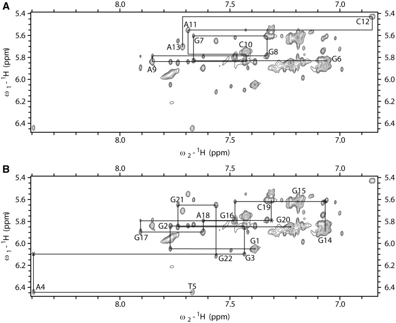
The aromatic–anomeric region of 2D NOESY spectrum (τm = 150 ms) of ODN. The lines connect aromatic H6/H8–sugar H1' NOE cross-peaks. Sequential connectivities for (A) G6-A13 segment and (B) G1-T5 and G14-G22 segments are shown separately to reduce cross-peaks’ overlap. Missing cross-peaks are designated with asterisk. 2D NOESY spectrum was recorded at 800 MHz, 25°C in 10% 2H2O, 50 mM KCl, 10 mM phosphate buffer with pH 6.8. Oligonucleotide concentration was 1.7 mM.
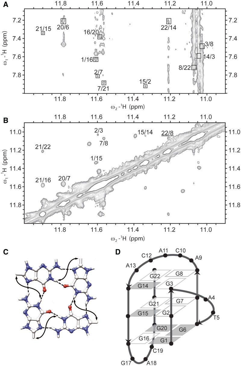
(A) The imino-aromatic region of 2D NOESY spectrum (τm = 150 ms). Cross-peaks between H1 and H8 protons of neighbouring guanine residues within the same quartet are designated with squares. (B) The imino–imino region of the same 2D NOESY spectrum. Cross-peak between G8 H1 and G22 H1 protons of neighbouring guanine residues in G-quartet is underlined. 2D NOESY spectrum was recorded at 800 MHz, 25°C in 10% 2H2O, 50 mM KCl, 10 mM phosphate buffer with pH 6.8. Oligonucleotide concentration was 1.7 mM. (C) Presentation of contacts between H1 and H8 protons (full arrows) of neighbouring guanine residues in G-quartet. (D) Schematic presentation of (3+1) topology of G-quadruplex adopted by ODN. Residues with syn and anti conformation along glycosidic bond are represented with grey and white rectangles, respectively.
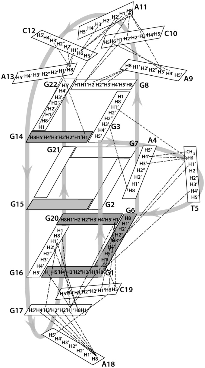
Schematic presentation of inter-residue NOE connectivities in loops of a G-quadruplex adopted by ODN. Residues with syn and anti conformation along glycosidic bond are represented with grey and white rectangles, respectively.
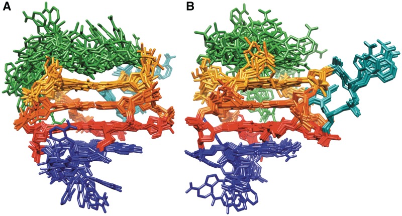
Superimposition of the 10 structures with the lowest energy and the smallest restraints violations. Figures in (A) and (B) are rotated with respect to one another to better represent superimposed structures. G1–G16–G20–G6, G2–G7–G21–G15 and G3–G8–G22–G14 quartets are shown in red, orange and yellow, respectively. Propeller (A4–T5), diagonal (A9–C10–A11–C12–A13) and edgewise (G17–A18–C19) loops are presented in cyan, green and blue, respectively.
Stacking interactions between (A) G1–G16–G20–G6 (blue) and G2–G7–G21–G15 (orange) quartets, and between (B) G2–G7–G21–G15 (orange) and G3–G8–G22–G14 (green) quartets.
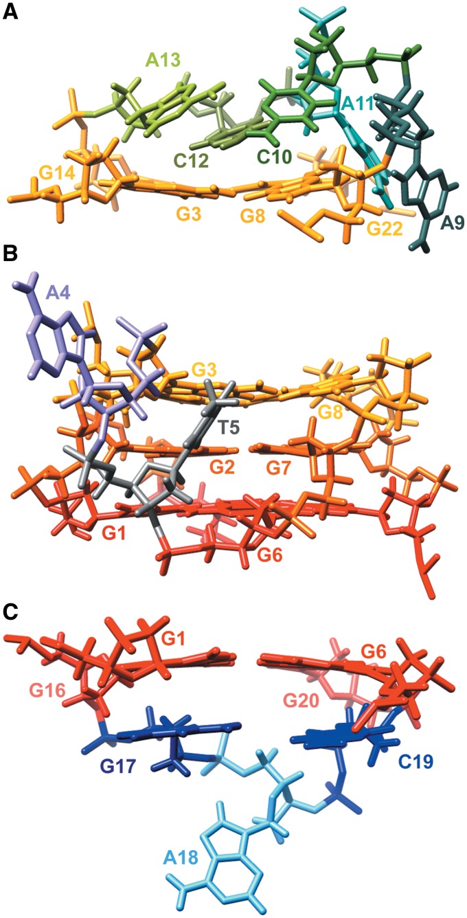
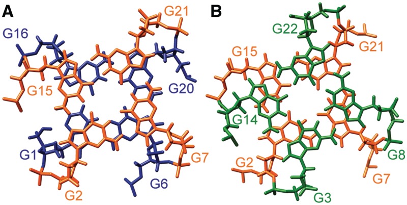
Position of loop residues in the G-quadruplex adopted by ODN. (A) Diagonal, (B) propeller and (C) edgewise loops in the representative structure, showing stacking of G17 and C19 on G1–G16–G20–G6 quartet, and stacking of C12 on G3–G8–G22–G14 quartet. G1–G16–G20–G6, G2–G7–G21–G15 and G3–G8–G22–G14 quartets are shown in red, orange and yellow, respectively.
References
-
- Wong HM, Payet L, Huppert JL. Function and targeting of G-quadruplexes. Curr. Opin. Mol. Ther. 2009;11:146–155. - PubMed
-
- Huppert JL. Structure, location and interactions of G-quadruplexes. FEBS J. 2010;277:3452–3458. - PubMed
-
- Verma A, Halder K, Halder R, Yadav VK, Rawal P, Thakur RK, Mohd F, Sharma A, Chowdhury S. Genome-wide computational and expression analyses reveal G-quadruplex DNA motifs as conserved cis-regulatory elements in human and related species. J. Med. Chem. 2008;51:5641–5649. - PubMed
Publication types
MeSH terms
Substances
Associated data
- Actions
LinkOut - more resources
Full Text Sources
Other Literature Sources
