. 2012;8(4):e1002645.
doi: 10.1371/journal.ppat.1002645. Epub 2012 Apr 19.

CD8+ T-cells expressing interferon gamma or perforin play antagonistic roles in heart injury in experimental Trypanosoma cruzi-elicited cardiomyopathy

Affiliations

CD8+ T-cells expressing interferon gamma or perforin play antagonistic roles in heart injury in experimental Trypanosoma cruzi-elicited cardiomyopathy

Jaline Coutinho Silverio et al. PLoS Pathog. 2012.

Abstract

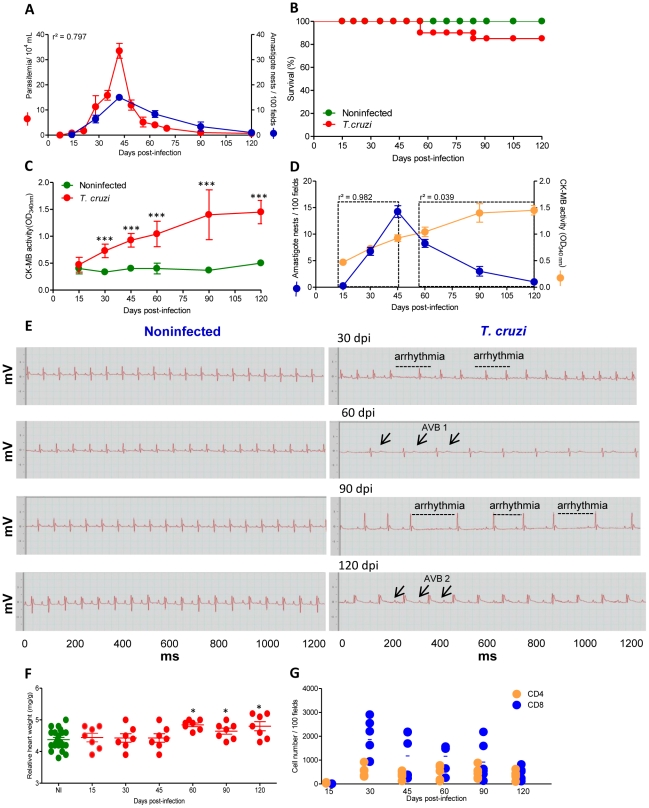
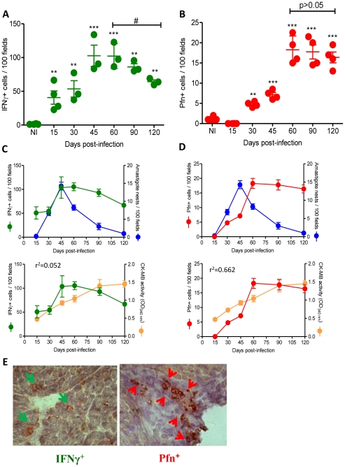
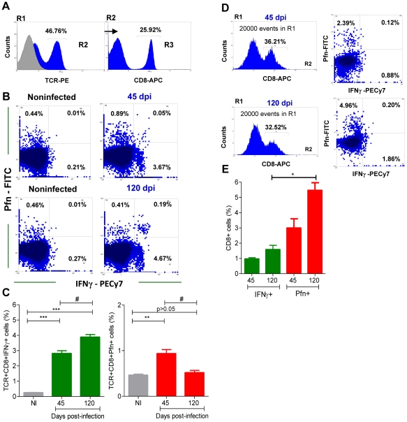
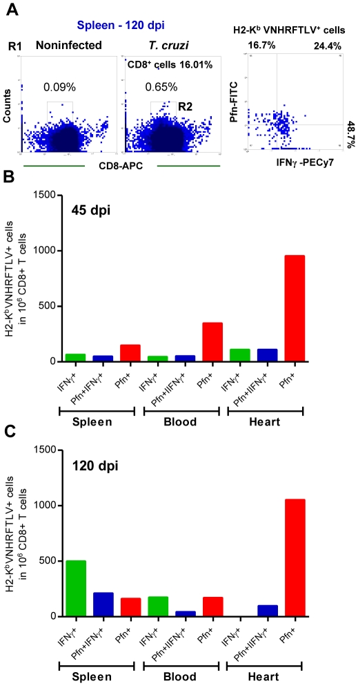
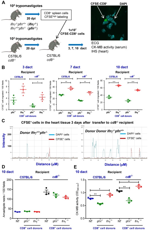
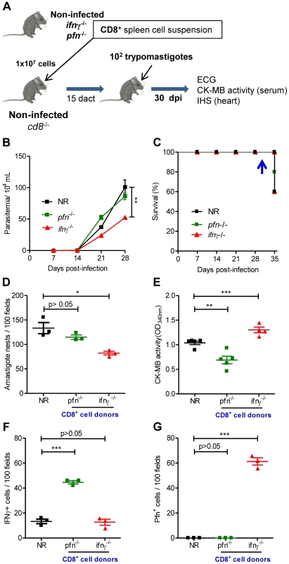
In Chagas disease, CD8(+) T-cells are critical for the control of Trypanosoma cruzi during acute infection. Conversely, CD8(+) T-cell accumulation in the myocardium during chronic infection may cause tissue injury leading to chronic chagasic cardiomyopathy (CCC). Here we explored the role of CD8(+) T-cells in T. cruzi-elicited heart injury in C57BL/6 mice infected with the Colombian strain. Cardiomyocyte lesion evaluated by creatine kinase-MB isoenzyme activity levels in the serum and electrical abnormalities revealed by electrocardiogram were not associated with the intensity of heart parasitism and myocarditis in the chronic infection. Further, there was no association between heart injury and systemic anti-T. cruzi CD8(+) T-cell capacity to produce interferon-gamma (IFNγ) and to perform specific cytotoxicity. Heart injury, however, paralleled accumulation of anti-T. cruzi cells in the cardiac tissue. In T. cruzi infection, most of the CD8(+) T-cells segregated into IFNγ(+) perforin (Pfn)(neg) or IFNγ(neg)Pfn(+) cell populations. Colonization of the cardiac tissue by anti-T. cruzi CD8(+)Pfn(+) cells paralleled the worsening of CCC. The adoptive cell transfer to T. cruzi-infected cd8(-/-) recipients showed that the CD8(+) cells from infected ifnγ(-/-)pfn(+/+) donors migrate towards the cardiac tissue to a greater extent and caused a more severe cardiomyocyte lesion than CD8(+) cells from ifnγ(+/+)pfn(-/-) donors. Moreover, the reconstitution of naïve cd8(-/-) mice with CD8(+) cells from naïve ifnγ(+/+)pfn(-/-) donors ameliorated T. cruzi-elicited heart injury paralleled IFNγ(+) cells accumulation, whereas reconstitution with CD8(+) cells from naïve ifnγ(-/-)pfn(+/+) donors led to an aggravation of the cardiomyocyte lesion, which was associated with the accumulation of Pfn(+) cells in the cardiac tissue. Our data support a possible antagonist effect of CD8(+)Pfn(+) and CD8(+)IFNγ(+) cells during CCC. CD8(+)IFNγ(+) cells may exert a beneficial role, whereas CD8(+)Pfn(+) may play a detrimental role in T. cruzi-elicited heart injury.


Conflict of interest statement

The authors have declared that no competing interests exist.

Figures

Figure 1
Figure 1. C57BL/6 mice infected with the Colombian strain of T. cruzi develop chronic cardiomyopathy.
Mice were infected with 100 bt of the Colombian strain of T. cruzi and parasitological and clinical parameters were evaluated. (A) Kinetics of parasitemia and cardiac parasitism. (B) Survival rate. (C) CK-MB activity levels in the serum of noninfected and T. cruzi-infected mice. (D) Positive correlation between cardiac tissue parasitism and CK-MB activity levels during the acute phase (r2 = 0.982) but not during the chronic phase (r2 = 0.039) of infection. (E) Representative ECG register segments of 1200 ms of sex- and age-matched noninfected (NI) controls and T. cruzi-infected C57BL/6 mice at 30, 45, 60, 90 and 120 dpi, showing arrhythmia and first and second degree atrioventricular block (AVB1, AVB2; arrows) in infected mice. (F) Relative heart weight (mg/g) of noninfected (NI, pool of three age-matched controls per analyzed point) and T. cruzi-infected C57BL/6 mice at 15, 30, 45, 60, 90 and 120 dpi. (G) Numbers of CD4+ and CD8+ cells in the cardiac tissue during acute and chronic T. cruzi infection were counted after immunohistochemistry staining. Each circle represents an individual mouse. These data represent three independent experiments. *, p<0.05, **, p<0.01, and ***, p<0.001 comparing NI controls and T. cruzi-infected mice.
Figure 2
Figure 2. CD8+ T-cells recognizing the VNHRFTLV ASP2 T. cruzi peptide are enriched in the cardiac tissue.
C57BL/6 mice were infected with 100 bt of the Colombian strain of T. cruzi and the anti-parasite immune response in the spleen and cardiac tissue was assessed. (A) Representative spots formed after stimulation of spleen cells from noninfected (NI) and T. cruzi-infected mice at 45 and 120 dpi with H-2Kb-resctricted VNHRFTLV peptide. The number of CD8+IFNγ+ as determined by ex vivo ELISpot were analyzed and compared with parasitemia and CK-MB activity levels in the serum. (B) Representative histogram profiles of in vivo cytotoxicity assay showing the specific lysis of H-2Kb-resctricted VNHRFTLV peptide-labeled CFSEhigh cells from NI controls and T. cruzi-infected C57BL/6 mice at 45 and 120 dpi. The frequencies of in vivo specific lysis of with H-2Kb -resctricted VNHRFTLV peptide-labeled target cells in T. cruzi-infected C57BL/6 mice at 15, 30, 45, 60, 90 and 120 dpi were analyzed and compared with parasitemia and CK-MB activity levels in the serum. The peak of parasitemia and the maximum CK-MB activity are highlighted (dotted line). Each circle and vertical lines represent the mean ± standard deviation (SD) of the studied group (5–7 animals/time point). These data represent three independent experiments. (C) Frequency of double-positive CD8+ H-2Kb /VNHRFTLV+ cells [R1 (SSCxFSC) gated] of spleen and heart of NI and T. cruzi-infected C57BL/6 mice at 40 dpi. (D) Frequencies of double-positive CD8+ H-2Kb /VNHRFTLV+ cells [R1 (SSCxFSC) gated] of spleen, peripheral blood and heart of NI and T. cruzi-infected C57BL/6 mice at 30, 45 and 120 dpi. Representative flow ctometry profiles and mean ± SD of two or three animals per group. Bars represent the mean of two or three pools of 5 mice per pool.
Figure 3
Figure 3. IFNγ+ and Pfn+ cells differentially invade the cardiac tissue of T. cruzi-infected C57BL/6 mice.
Mice were infected with 100 bt of the Colombian strain of T. cruzi and the presence of IFNγ+ and Pfn+ cells in the cardiac tissue was evaluated by IHS. (A) Numbers of IFNγ+ cells infiltrating the cardiac tissue at 15, 30, 45, 60, 90 and 120 dpi. (B) Numbers of Pfn+ cells infiltrating the cardiac tissue at 15, 30, 45, 60, 90 and 120 dpi. (C) Relationship between IFNγ+ cells infiltrating the cardiac tissue and heart parasitism or CK-MB activity in serum. (D) Relationship between Pfn+ cells infiltrating the cardiac tissue and heart parasitism or CK-MB activity in the serum. (E) Immunohistochemistry staining of IFNγ+ (green arrows) and Pfn+ (red arrows) cells infiltrating the cardiac tissue at 60 dpi. In (A) and (B), each circle represents an individual mouse. In (C) and (D), each circle represents the mean ± SD of the studied group (5–7 animals/time point). These data represent three independent experiments. *, p<0.05; **, p<0.01; and ***, p<0.001, comparing noninfected (NI) controls and T. cruzi-infected mice. #, p<0.05, comparing T. cruzi-infected mice at 60 and 120 dpi.
Figure 4
Figure 4. Segregation of CD8+ T-cells into IFNγ+ and Pfn+ populations in T. cruzi infection.
C57BL/6 mice were infected with 100 blood trypomastigotes of the Colombian strain of T. cruzi and the expression of IFNγ+ and Pfn+ by CD8+ cells in the peripheral blood and cardiac tissue was evaluated. (A) Representative histograms of flow cytometry analysis of peripheral blood CD8+ T-cells [R1 (SSCxFSC)/R2 (TCR)/R3 (CD8) gated] that were analyzed for IFNγ and Pfn expression in T. cruzi-infected mice (120 dpi). (B) Representative dot plots of flow cytometry analysis of peripheral blood CD8+ T-cells (R1/R2/R3 gated) expressing IFNγ and Pfn at 45 and 120 dpi. (C) Frequency of double-stained CD8+IFNγ+ and CD8+Pfn+ peripheral blood T-cells cells (R1/R2/R3 gated) at 45 and 120 dpi. (D) Representative histograms and dot plots of flow cytometry analysis of heart infiltrating CD8+ cells [R1 (SSCxFSC)/R2 (CD8) gated] expressing IFNγ and Pfn at 45 and 120 dpi. (E) Frequency of double-stained CD8+IFNγ+ and CD8+Pfn+ of heart infiltrating cells (R1/R2 gated) at 45 and 120 dpi. In (C) Bars represent the mean ± SD of five to eight mice per group. In (E) Bars represent the mean of two or three pools of 5 mice per group. These data represent two or three independent experiments. *, p<0.05; **, p<0.01; and ***, p<0.001, comparing noninfected (NI) controls and T. cruzi-infected mice. # P<0.05, comparing T. cruzi-infected mice at 45 and 120 dpi.
Figure 5
Figure 5. Differential compartmentalization of anti-T. cruzi CD8+ T-cells expressing IFNγ and Pfn.
C57BL/6 mice were infected with 100 bt of the Colombian strain of T. cruzi and presence of CD8+ H-2Kb /VNHRFTLV+ T-cells expressing IFNγ+ and Pfn+ in the spleen, peripheral blood and in the cardiac tissue was evaluated. (A) Representative dot plots of double-positive CD8+ H-2Kb /VNHRFTLV+ T-cells (R1 gated) in the spleen of NI and T. cruzi-infected C57BL/6 mice at 120 dpi. Frequencies of CD8+ H-2Kb /VNHRFTLV+ T-cells (R2 gated) expressing IFNγ, Pfn or coexpressing IFNγ and Pfn in the spleen, peripheral blood and cardiac tissue of NI and T. cruzi-infected C57BL/6 mice at 45 (B) and 120 dpi (C). Bars represent the means of two or three pools of 5 mice per group. These data represent two independent experiments.
Figure 6
Figure 6. CD8+ cells from ifnγ −/− pfn +/+ and ifnγ +/+ pfn −/− infected mice differentially migrate to the cardiac tissue.
(A) Experimental scheme showing the infection of donors and recipients mice by T. cruzi, isolation of CD8+ cells from ifnγ −/− pfn +/+ and ifnγ +/+ pfn −/− infected donors (20 dpi) by magnetic beads, CFSEhigh labeling, adoptive transfer of CFSE+CD8+ cells to C57BL/6 and cd8 −/− infected recipients (20 dpi) and analysis at 3, 7 and 10 days after cell transfer (dact). The colonization of the cardiac tissue by CFSE+ cells, heart parasitism and CK-MB activity levels in the serum were evaluated. (B) Detection of CFSE+CD8+ cells in cardiac tissue sections at 3, 7 and 10 dact to T. cruzi-infected C57BL/6 and cd8 −/− recipients. (C) Graphics showing the confocal analysis of the intensity of fluorescence of CFSE+CD8+ cells present in the myocardium of cardiac tissue sections at 3 dact to T. cruzi-infected cd8 −/− recipients. DAPI was used to reveal the nucleus of the cells of the recipient mice. (D) Number of amastigote nests in 100 microscopic fields of cardiac tissue from C57BL/6 and cd8 −/− infected mice that were non-transferred (NT) or transferred with CD8+ cells from ifnγ −/− pfn +/+ and ifnγ +/+ pfn −/− donors, at 10 dact. (E) Cardiomyocyte lesions were evaluated at 10 dact by measuring the CK-MB activity in serum samples from C57BL/6 and cd8 −/− infected mice that were non-transferred (NT) or transferred with CD8+ cells from ifnδ −/− pfn +/+ and ifnγ +/+ pfn −/− donors. Each symbol represents an individual mouse. These data represent three independent experiments. *, p<0.05; **, p<0.01; and ***, p<0.001, comparing C57BL/6 and cd8 −/− infected mice that were non-transferred and transferred with CD8+ cells from ifnγ −/− pfn +/+ and ifnγ +/+ pfn −/− donors.
Figure 7
Figure 7. Distinct migratory behavior and effector function of CD8+IFNγ+ and CD8+Pfn+ T-cells in T. cruzi infection.
(A) Experimental scheme of CD8+ cell isolation from noninfected naïve ifnγ −/− pfn +/+ and ifnγ +/+ pfn −/− donors, in vivo reconstitution of the CD8+ cell compartment of naïve cd8 −/− mice, infection with 100 bt of the Colombian strain at 15 days after cell transfer (dact) and analysis at 30 days post-infection. Parasitemia, survival rate, cardiac parasitism and CK-MB activity levels in the serum and colonization of the cardiac tissue by IFNγ+ and Pfn+ cells were evaluated. (B) Parasitemia and (C) survival curve of cd8 −/− mice non-reconstituted (NR) or reconstituted with CD8+ cells from ifnγ +/+ pfn −/− and ifnγ −/− pfn +/+ donors. In independent experiments, the animals were analyzed at 30 dpi when 100% of the mice in all the experimental groups were alive (arrow). (D) Number of amastigote nests in 100 microscopic fields of cardiac tissue sections. (E) Cardiomyocyte lesion evaluated by CK-MB activity in serum samples. The number of (F) IFNγ+ and (G) Pfn+ cells in 100 microscopic fields of cardiac tissue sections from mice non-reconstituted (NR) or reconstituted with CD8+ cells from ifnγ +/+ pfn −/− and ifnγ −/− pfn +/+ donors, at 30 dpi. Each symbol represents an individual mouse. These data represent three independent experiments. *, p<0.05; **, p<0.01; and ***, p<0.001, comparing cd8 −/− infected mice non-reconstituted and reconstituted with CD8+ cells from ifnγ −/− pfn +/+ and ifnγ +/+ pfn −/− donors.

References

    1. Lannes-Vieira J, de Araújo-Jorge TC, Soeiro Mde N, Gadelha P, Corrêa-Oliveira R. The centennial of the discovery of Chagas disease: facing the current challenges. PLoS Negl Trop Dis. 2010;4:e645. - PMC - PubMed
    1. Rassi A, Jr, Rassi A, Marin-Neto JA. Chagas disease. Lancet. 2010;375:1388–13402. - PubMed
    1. Dutra WO, Menezes CA, Villani FN, da Costa GC, da Silveira AB, et al. Cellular and genetic mechanisms involved in the generation of protective and pathogenic immune responses in human Chagas disease. Mem Inst Oswaldo Cruz. 2009;104:208–218. - PMC - PubMed
    1. Padilla AM, Bustamante JM, Tarleton RL. CD8+ T cells in Trypanosoma cruzi infection. Curr Opin Immunol. 2009;21:385–390. - PMC - PubMed
    1. Higuchi Mde L, Gutierrez PS, Aiello VD, Palomino S, Bocchi E, et al. Immunohistochemical characterization of infiltrating cells in human chronic chagasic myocarditis: comparison with myocardial rejection process. Virchows Arch A Pathol Anat Histopathol. 1993;423:157–160. - PubMed

Publication types

MeSH terms