. 2012 Feb;1(2):96-109.
doi: 10.5966/sctm.2011-0057.

Generation of retinal pigment epithelial cells from small molecules and OCT4 reprogrammed human induced pluripotent stem cells

Affiliations

Generation of retinal pigment epithelial cells from small molecules and OCT4 reprogrammed human induced pluripotent stem cells

Tim U Krohne et al. Stem Cells Transl Med. 2012 Feb.

Abstract

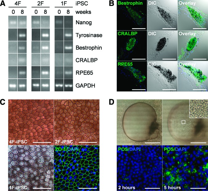
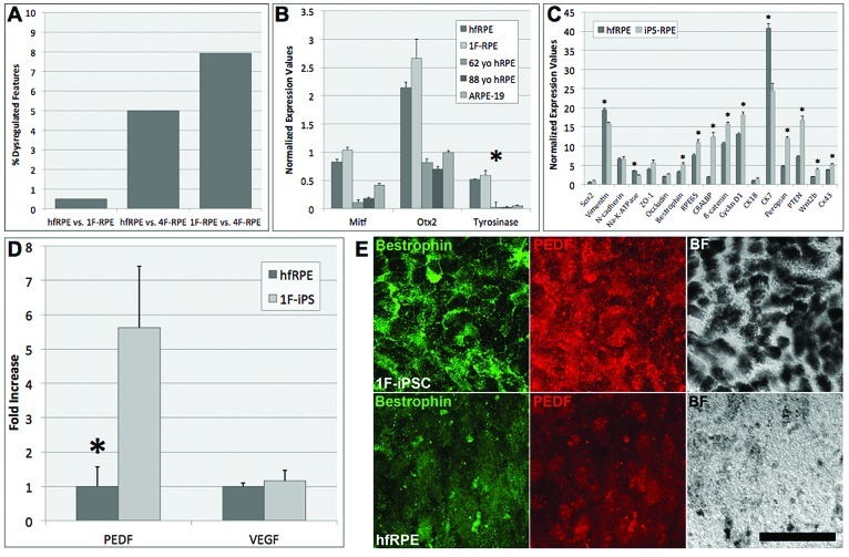
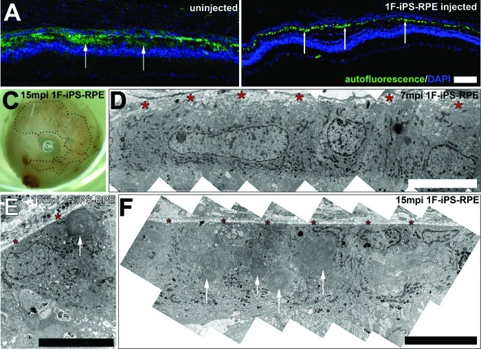
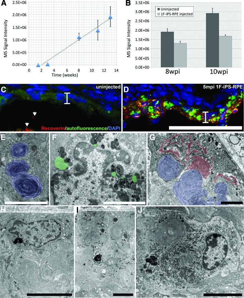
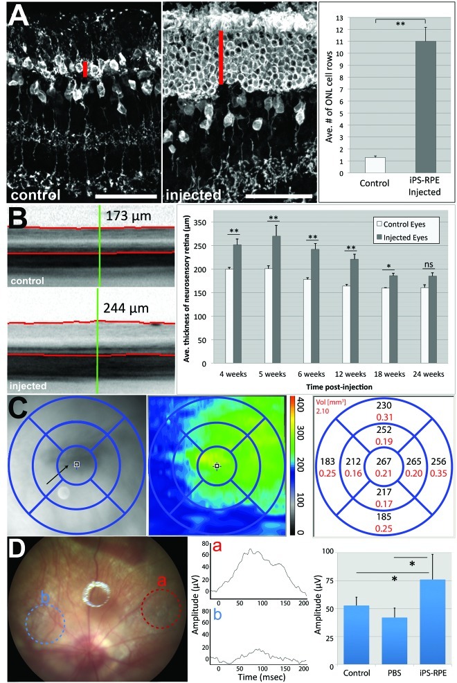
Autologous retinal pigment epithelium (RPE) grafts derived from induced pluripotent stem cells (iPSCs) may be used to cure blinding diseases in which RPE dysfunction results in photoreceptor degeneration. Four-, two-, and one-factor-derived iPSCs (4F-, 2F-, and 1F-iPSCs, respectively) were differentiated into fully functional cuboidal pigmented cells in polarized monolayers that express RPE-specific markers. 1F-iPSCs-RPE (1F-iPS-RPE) strongly resembles primary human fetal RPE (hfRPE) based on proteomic and untargeted metabolomic analyses, and using novel in vivo imaging technology coupled with electroretinography, we demonstrated that 1F-iPS-RPE mediate anatomical and functional rescue of photoreceptors after transplantation in an animal model of RPE-mediated retinal degeneration. 1F-iPS0RPE cells were injected subretinally as a suspension and formed a monolayer dispersed between host RPE cells. Furthermore, 1F-iPS-RPE do not simply provide trophic support to rescue photoreceptors as previously speculated but actually phagocytose photoreceptor outer segments in vivo and maintain visual cycling. Thus, 1f-iPS-RPE grafts may be superior to conventional iPS-RPE for clinical use because 1F-IPS-RPE closely resemble hfRPE, mediate anatomical and functional photoreceptor rescue in vivo, and are generated using a reduced number of potentially oncogenic reprogramming factors.

Keywords: Retinal pigment epithelium; differentiation; induced pluripotent stem cells; small molecules.


Figures

Figure 1.
Figure 1.
(A): mRNA expression was analyzed in 4F-, 2F-, and 1F-iPSCs at the beginning and end of 8 weeks of directed differentiation. Expression of the pluripotency marker Nanog decreased with differentiation but remained detectable in 4F-iPSCs that were transfected with Nanog during reprogramming. Expression of the melanogenesis marker tyrosinase was induced during differentiation, and its expression correlated closely with development of cellular pigmentation. RPE terminal differentiation markers bestrophin, CRALBP, and RPE65 were minimally expressed in undifferentiated iPSCs but strongly expressed in differentiated cells. GAPDH served as loading control. (B): Immunohistochemical analysis of differentiated 4F-, 2F-, and 1F-iPS-RPE confirmed protein expression of terminal RPE differentiation markers and a clear localization in cells also expressing pigment (representative results of 2F-iPS-RPE shown). (C): Isolation and expansion of pigmented cell clusters from differentiated 4F-, 2F-, and 1F-iPS-RPE yielded homogenous cultures of cells with morphological features characteristic of RPE cells, such as monolayer growth, hexagonal shape, and pronounced pigmentation. Expression and localization of the tight junction protein ZO-1 was detectable in 4F-, 2F-, and 1F-iPS-RPE (representative results of 4F-iPS-RPE shown). (D): iPS-RPE exhibited RPE functionality in vitro including directional fluid transport and photoreceptor outer segment (POS) phagocytosis. Barrier properties of the cellular monolayer in combination with apical-to-basolateral fluid transport resulted in progressive formation of fluid-filled domes in 4F-, 2F-, and 1F-iPS-RPE cultures (top panels, representative results of 4F-iPS-RPE). Furthermore, POS phagocytosis was detectable in 4F-, 2F-, and 1F-iPS-RPE (bottom panels, representative results of 2F-iPS-RPE). Two hours after addition of fluorescein-5-isothiocyanate-labeled POS to iPSC-RPE cultures, the POS were bound but not yet internalized. The increase in detectable POS after 5 hours represents POS internalization. Scale bars = 100 μm (B), 50 μm (C), 1000 μm (D, top), and 50 μm (D, bottom). Abbreviations: 1F, one-factor; 2F, two-factor; 4F, four-factor; DAPI, 4′,6-diamidino-2-phenylindole; DIC, Differential interference contrast; GAPDH, glyceraldehyde-3-phosphate dehydrogenase; iPSC, induced pluripotent stem cell; RPE, retinal pigment epithelium.
Figure 2.
Figure 2.
(A): The percentage of dysregulated metabolic features (fold change ≥2 with high statistical significance, p ≤ 0.001) was compared between hfRPE, 1F-iPS-RPE (1F-iPS-2), and 4F-iPS-RPE. Nominal disparities were observed between hfRPE and 1F-iPS-RPE (0.5%; 82 of 15,651 total features). A 10-fold higher percentage of dysregulated features was observed between hfRPE and 4F-RPE although the overall difference was still low (5.0%; 691 of 13,853 total features). Few differences were observed between 1F-iPS-RPE and 4F-iPS-RPE (7.9%; 1,320 of 16,682 total features); however, both 1F and 4F seemed to be more similar to hfRPE than each other. (B): In-cell Western analyses comparing normalized protein expression values between hfRPE, 1F-iPS-RPE (1F-iPS-2), 4F-iPS-RPE, 62-year-old hRPE, 88-year-old hRPE, and ARPE-19 cells revealed differences in expression levels of Mitf, Otx2, and tyrosinase. hfRPE and 1F-iPS-RPE were consistently the most similar of all the cell types examined. One-way analysis of variance tests revealed that the differences in tyrosinase expression between the groups were statistically significant (p = .2). (C): Direct comparison of the normalized expression values collected from the in-cell Western analysis in hfRPE and 1F-iPS-RPE for the proteins listed on the x-axis. (D): A quantitative comparison of PEDF and VEGF secretion by hfRPE and 1F-iPS-RPE (1F-iPSC-2) cells was performed using sandwich enzyme-linked immunosorbent assays. The results were plotted as fold increase above hfRPE levels. A 5.6-fold (p = .019) difference in secreted PEDF was detected between the hfRPE and 1F-iPS-RPE. No significant difference in secreted VEGF was detected (1.2-fold, p = .68) between the two cell types. (E): Immunocytochemistry was performed on 1F-iPSC-2 and hfRPE grown on Transwell filters. Bestrophin labeling (green) in 1F-iPSC-RPE staining marks the well-defined basolateral cell membranes but was diffusely expressed in hfRPE. PEDF (red) was more readily detectable in 1F-iPS-RPE than hfRPE, and much more pigment was synthesized in 1F-iPSC-RPE cultures than in hfRPE, as observed in the BF images. Collectively, these observations demonstrate that 1F-iPSC-RPE cells strongly resemble hfRPE but are in an advanced differentiation state. Errors bars in all panels represent SEM values. Scale bar = 50 μm (E). Abbreviations: 1F, one-factor; 4F, four-factor; BF, bright field; hfRPE, human fetal retinal pigment epithelium; hRPE, human retinal pigment epithelium; iPS, induced pluripotent stem; iPSC, induced pluripotent stem cell; RPE, retinal pigment epithelium; VEGF, vascular endothelial growth factor; yo, year old.
Figure 3.
Figure 3.
(A): Autofluorescence in uninjected Royal College of Surgeons (RCS) rat eyes was detected in a broad diffuse region correlating with the subretinal space. (B): In contralateral eyes injected with 1F-iPS-RPE (1F-iPS-2), the autofluorescence pattern was punctate and limited to the RPE cell layer. (C): Implanted pigmented 1F-iPS-RPE cells could be observed in albino RCS rat eyes after enucleation. (D): The implanted 1F-iPS-RPE cells incorporated into the RPE layer and were correctly polarized. Red asterisks mark Bruch's membrane. (E, F): Implanted cells could also be detected 15 months after implantation. Arrows point to debris accumulation in the implanted 1F-iPS-RPE cells. Scale bars = 100 μm (A, B) and 10 μm (D–F). Abbreviations: 1F, one-factor; iPS, induced pluripotent stem; mpi, months postinjection; RPE, retinal pigment epithelium.
Figure 4.
Figure 4.
1F-iPS-RPE (clone 1F-iPS-2) phagocytosed photoreceptor outer segment debris and reduced the levels of A2E accumulation in Royal College of Surgeons (RCS) rat eyes. (A): A2E accumulation in RCS rat as detected by liquid chromatography-mass spectrometry. (B): At 8 (n = 10) and 10 (n = 6) wpi with 1F-iPS-RPE, the injected eyes accumulated less A2E (66.9%, p = .03, and 71.2%, p = .006 respectively). (C): In uninjected RCS rat eyes, nominal and diffuse autofluorescence and no recoverin (arrowheads) was detected in RPE cells. (D): Conversely, granular and highly autofluorescent lipofuscin and recoverin-positive material was detected in the RCS rat eyes after 1F-iPS-RPE cell implantation (the RPE monolayer is marked with a bracket [C, D]). (E): 1F-iPS-RPE phagocytosed material morphologically resembling photoreceptor outer segments 7 mpi. (F, G): 15 mpi, what appeared to be lipofuscin (green) and extracellular deposits (red) accumulated in 1F-iPS-RPE cells. (H–J): Host RPE (H), implanted 1F-iPS-RPE (I), and a host macrophage (J). Scale bars = 50 μm (C, D), 2 μm in (E–G), and 5 μm (H–J). Abbreviations: 1F, one-factor; DAPI, 4′,6-diamidino-2-phenylindole; iPS, induced pluripotent stem; mpi, months postinjection; MS, mass spectrometry; RPE, retinal pigment epithelium; wpi, weeks postinjection.
Figure 5.
Figure 5.
(A): Immunohistochemistry analyses were performed 6 weeks after transplantation of 1F-iPS-RPE (clone 1F-iPSC-2) in the subretinal space of RCS rats. Recoverin staining labeled photoreceptors (marked with a red line) and some bipolar cells. Note that at this age (9 weeks old, or 6 weeks postinjection), the ONL was virtually depleted in the uninjected control eyes, but a significant number of photoreceptors had been preserved in the ONL of the eyes injected with iPS-RPE. The effects were quantified by counting the layers of nuclei in the ONL (right panel). (B): In vivo optical coherence tomography (OCT) images from 9-week-old control and injected RCS rat eyes across several weeks were taken and analyzed using a spectral-domain OCT system (Spectralis, Heidelberg Engineering). The red lines mark the inner layer of the retina (top) and the photoreceptor outer segment (POS)/RPE interface (bottom). The green reference line marks the point where was the thickness measurements were calculated between the red lines. These measurements were averaged and plotted (right panel). (C): Regions of the eyes where 1F-iPS-RPE cells had successfully integrated were analyzed as described above. For this analysis, 25 images were taken across the region marked with a grid. Thickness measurements from this region are shown as a heat map (middle panel) and as averaged values (right panel). Note that the thickest regions correlate with the region in the fundus image where RPE cells were detected (the RPE cells in albino RCS rats do not synthesize melanin pigment). (D): Fundus image of an RCS rat with 1F-iPS-RPE cells implanted (n = 4; representative image and traces are shown). The red circle (a) marks where the light beam was aimed in the eye over the 1F-iPS-RPE implanted region; the resulting electrical activity is shown in the trace labeled (a) in the middle panel and quantified in the right panel. The blue circle (b) marks where no cells were implanted; a significantly lower response was detected from this region (shown in the middle panel and quantified in the right panel). Focal electroretinography was used to test the electrical activity in RCS rat eyes injected with PBS. Activity in these eyes was similar to that of uninjected controls. Abbreviations: Ave., average; iPS, induced pluripotent stem; ONL, outer nuclear layer; PBS, phosphate-buffered saline; RPE, retinal pigment epithelium.

References

    1. Yu J, Vodyanik MA, Smuga-Otto K, et al. Induced pluripotent stem cell lines derived from human somatic cells. Science. 2007;318:1917–1920. - PubMed
    1. Takahashi K, Tanabe K, Ohnuki M, et al. Induction of pluripotent stem cells from adult human fibroblasts by defined factors. Cell. 2007;131:861–872. - PubMed
    1. Koch P, Kokaia Z, Lindvall O, et al. Emerging concepts in neural stem cell research: Autologous repair and cell-based disease modelling. Lancet Neurol. 2009;8:819–829. - PubMed
    1. Aoi T, Yae K, Nakagawa M, et al. Generation of pluripotent stem cells from adult mouse liver and stomach cells. Science. 2008;321:699–702. - PubMed
    1. Okita K, Ichisaka T, Yamanaka S. Generation of germline-competent induced pluripotent stem cells. Nature. 2007;448:313–317. - PubMed

Publication types

MeSH terms