

An FMRI study of auditory orienting and inhibition of return in pediatric mild traumatic brain injury
- PMID: 22533632
- PMCID: PMC3419846
- DOI: 10.1089/neu.2012.2395
An FMRI study of auditory orienting and inhibition of return in pediatric mild traumatic brain injury
Abstract
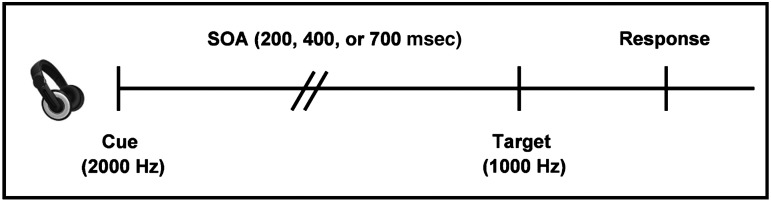
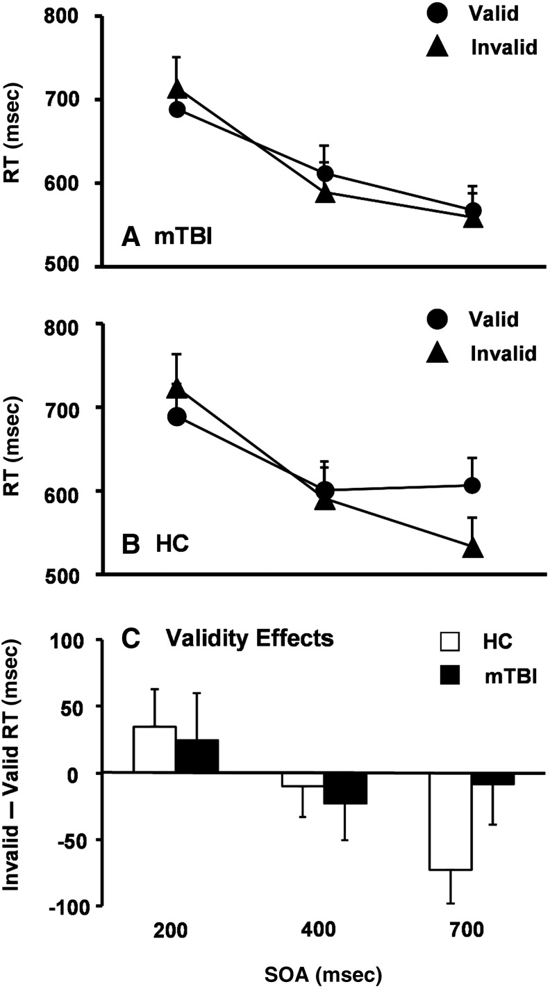
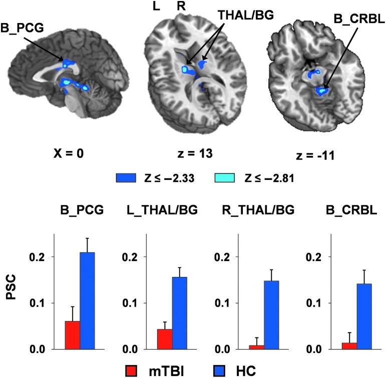
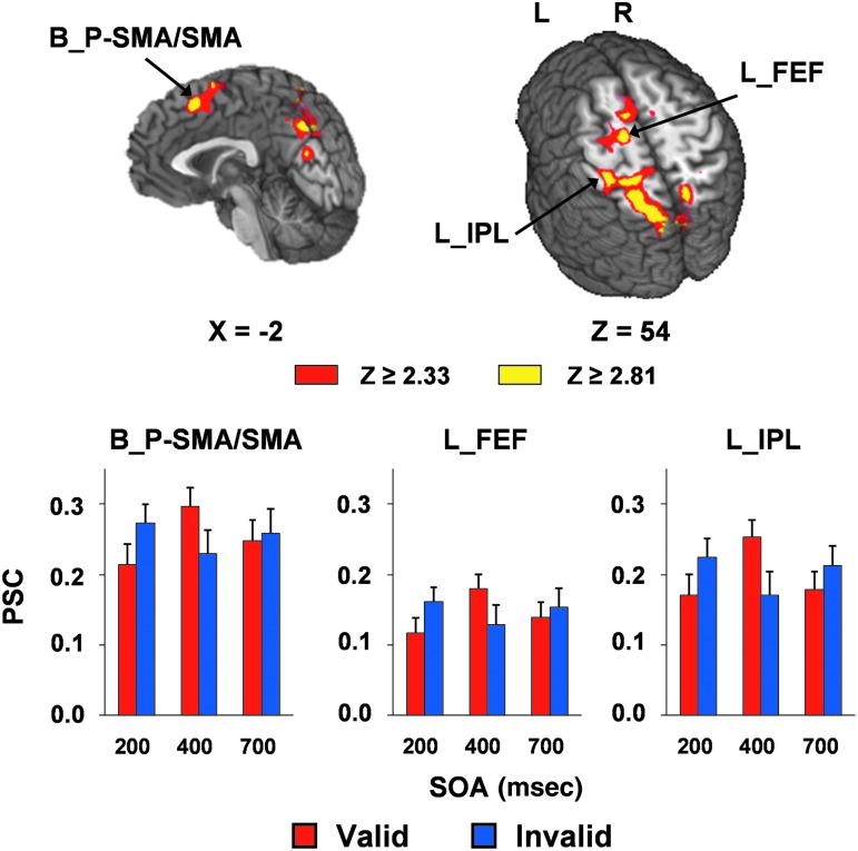
Studies in adult mild traumatic brain injury (mTBI) have shown that two key measures of attention, spatial reorienting and inhibition of return (IOR), are impaired during the first few weeks of injury. However, it is currently unknown whether similar deficits exist following pediatric mTBI. The current study used functional magnetic resonance imaging (fMRI) to investigate the effects of semi-acute mTBI (<3 weeks post-injury) on auditory orienting in 14 pediatric mTBI patients (age 13.50±1.83 years; education: 6.86±1.88 years), and 14 healthy controls (age 13.29±2.09 years; education: 7.21±2.08 years), matched for age and years of education. The results indicated that patients with mTBI showed subtle (i.e., moderate effect sizes) but non-significant deficits on formal neuropsychological testing and during IOR. In contrast, functional imaging results indicated that patients with mTBI demonstrated significantly decreased activation within the bilateral posterior cingulate gyrus, thalamus, basal ganglia, midbrain nuclei, and cerebellum. The spatial topography of hypoactivation was very similar to our previous study in adults, suggesting that subcortical structures may be particularly affected by the initial biomechanical forces in mTBI. Current results also suggest that fMRI may be a more sensitive tool for identifying semi-acute effects of mTBI than the procedures currently used in clinical practice, such as neuropsychological testing and structural scans. fMRI findings could potentially serve as a biomarker for measuring the subtle injury caused by mTBI, and documenting the course of recovery.
Figures
References
-
- Adelson P.D. Kochanek P.M. Head injury in children. J. Child Neurol. 1998;13:2–15. - PubMed
-
- Allen D.N. Leany B.D. Thaler N.S. Cross C. Sutton G.P. Mayfield J. Memory and attention profiles in pediatric traumatic brain injury. Arch. Clin. Neuropsychol. 2010;25:618–633. - PubMed
-
- Anderson V. Catroppa C. Morse S. Haritou F. Rosenfeld J. Outcome from mild head injury in young children: a prospective study. J. Clin. Exp. Neuropsychol. 2001;23:705–717. - PubMed
-
- Anderson V. Catroppa C. Morse S. Haritou F. Rosenfeld J. Attentional and processing skills following traumatic brain injury in early childhood. Brain Inj. 2005;19:699–710. - PubMed
-
- Arrington C.M. Carr T.H. Mayer A.R. Rao S.M. Neural mechanisms of visual attention: object-based selection of a region in space. J. Cogn. Neurosci. 2000;12(Suppl. 2):106–117. - PubMed
Publication types
MeSH terms
Grants and funding
LinkOut - more resources
Full Text Sources
