. 2012 Apr 25:9:76.
doi: 10.1186/1742-2094-9-76.

Neuroprotection by inhibiting the c-Jun N-terminal kinase pathway after cerebral ischemia occurs independently of interleukin-6 and keratinocyte-derived chemokine (KC/CXCL1) secretion

Affiliations

Neuroprotection by inhibiting the c-Jun N-terminal kinase pathway after cerebral ischemia occurs independently of interleukin-6 and keratinocyte-derived chemokine (KC/CXCL1) secretion

Corinne Benakis et al. J Neuroinflammation. .

Abstract

Background: Cerebral ischemia is associated with the activation of glial cells, infiltration of leukocytes and an increase in inflammatory mediators in the ischemic brain and systemic circulation. How this inflammatory response influences lesion size and neurological outcome remains unclear. D-JNKI1, an inhibitor of the c-Jun N-terminal kinase pathway, is strongly neuroprotective in animal models of stroke. Intriguingly, the protection mediated by D-JNKI1 is high even with intravenous administration at very low doses with undetectable drug levels in the brain, pointing to a systemic mode of action, perhaps on inflammation.

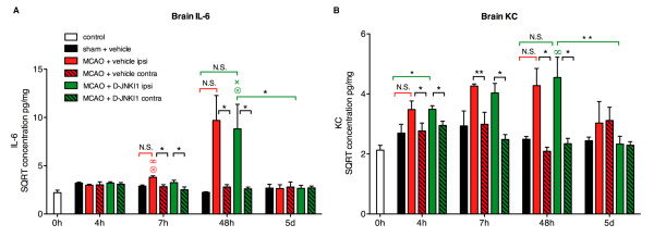
Findings: We evaluated whether D-JNKI1, administered intravenously 3 h after the onset of middle cerebral artery occlusion (MCAO), modulates secretion of the inflammatory mediators interleukin-6 and keratinocyte-derived chemokine in the plasma and from the spleen and brain at several time points after MCAO. We found an early release of both mediators in the systemic circulation followed by an increase in the brain and went on to show a later systemic increase in vehicle-treated mice. Release of interleukin-6 and keratinocyte-derived chemokine from the spleen of mice with MCAO was not significantly different from sham mice. Interestingly, the secretion of these inflammatory mediators was not altered in the systemic circulation or brain after successful neuroprotection with D-JNKI1.

Conclusions: We demonstrate that neuroprotection with D-JNKI1 after experimental cerebral ischemia is independent of systemic and brain release of interleukin-6 and keratinocyte-derived chemokine. Furthermore, our findings suggest that the early systemic release of interleukin-6 and keratinocyte-derived chemokine may not necessarily predict an unfavorable outcome in this model.


Figures

Figure 1
Figure 1
Two peaks of interleukin-6 and keratinocyte-derived chemokine release into the plasma after middle cerebral artery occlusion. Square root-transformed (SQRT) IL-6 and KC plasma concentrations at different time points in control mice, sham and MCAO mice treated with vehicle or D-JNKI1. (A) In sham mice, IL-6 did not differ from control mice at any time point. Plasma IL-6 was induced 4 h and 7 h after MCAO+vehicle at a significantly higher level than sham mice. At 24 h (data not shown) and 48 h, IL-6 concentrations in MCAO+vehicle mice returned to basal level and increased again 5 d after the occlusion (sham versus MCAO+vehicle: P = 0.054). The plasma IL-6 concentration in MCAO+D-JNKI1 mice was slightly reduced at 4 h and 5 d but not statistically different from MCAO+vehicle mice (4 h: MCAO+vehicle versus MCAO+D-JNKI1: P = 0.26; 5 d: MCAO+vehicle versus MCAO+D-JNKI1: P = 0.27). Seven hours after MCAO, IL-6 concentration in D-JNKI1-treated mice was significantly higher compared with sham mice. (B) Plasma KC was induced early in MCAO+vehicle mice compared to sham mice and decreased from 24 h (data not shown) onwards. Five days after MCAO, KC concentration was again significantly higher than in sham mice (sham versus MCAO+vehicle: P = 0.03). KC concentration in MCAO+D-JNKI1 mice was similar to MCAO+vehicle mice, without any statistical difference between the two groups. In the sham group, KC concentrations at 4 h, 7 and 24 h (data not shown) were significantly higher than in controls. Control mice: n = 7; sham mice: n = 5 to 8, except at 24 h, n = 3; MCAO mice: n = 6 to 10. *P <0.05, **P <0.01 indicate a significant difference between MCAO and sham mice. Results are presented as mean ± SEM. When placed over a data point, ∞, ⊗ and × indicate a significant .difference with concentrations in the same group at 0 h, 4 h and 7 h, respectively.
Figure 2
Figure 2
Release of interleukin-6 and keratinocyte-derived chemokine from the spleen after middle cerebral artery occlusion. Concentrations of IL-6 and KC were measured in the supernatants of spleen slice cultures and normalized to total protein concentrations. The graphs show the square root (SQRT) of (A) IL-6 and (B) KC concentrations in pg/mg at different time points from control (n = 7), sham (n = 4 to 8), MCAO+vehicle (n = 6 to 10) and MCAO+D-JNKI1 (n = 5 to 8) mice. IL-6 release from the spleen increased slightly, though not significantly, at 4 h in both MCAO+vehicle and MCAO+D-JNKI1 mice. Overall, there is no significant change in IL-6 and KC in spleen supernatants in any of the four groups tested. Results are represented as mean ± SEM.
Figure 3
Figure 3
Release of interleukin-6 and keratinocyte-derived chemokine from the brain after middle cerebral artery occlusion. Concentrations of IL-6 and KC were measured in the supernatants of brain slice cultures and normalized to total protein concentrations. The graphs show the square root (SQRT) of IL-6 and KC concentrations in pg/mg at different time points in control, sham and MCAO mice. In sham mice, the cerebral release of IL-6 and KC did not differ from control mice at any time point. (A) Secretion of IL-6 from the brain tended to increase at 24 h (sham versus MCAO+vehicle: P = 0.06, data not shown) with a significant difference between sham and MCAO+vehicle 48 h after the occlusion. A slight, non-significant reduction of IL-6 release from the brain in mice treated with D-JNKI1 was seen at 5 d (MCAO+vehicle versus MCAO+D-JNKI1: P = 0.25). There was no significant difference of brain IL-6 release in D-JNKI1- and vehicle-treated mice at any time point. In MCAO + D-JNKI1 mice, IL-6 concentrations at 48 h remained significantly higher than in controls. (B) KC release in MCAO + vehicle mice started to increase significantly at 7 h compared to early time points (0 h and 4 h) and remained elevated at late time points. The brain KC release increased slightly, though not significantly, at 24 h (P = 0.19, data not shown) and 48 h (P = 0.08) after MCAO compared with sham mice. KC concentrations in MCAO + D-JNKI1 mice followed the same temporal profile as in MCAO + vehicle mice. Control mice: n = 7; sham mice: n = 5 to 8, except at 24 h, n = 3; MCAO mice: n = 6 to 10. *P <0.05 indicates a significant difference in MCAO mice and sham mice. Results are represented as mean ± SEM. When placed over a data point, ∞ and ⊗ indicate a significant difference with concentrations in the same group at 0 h and 4 h, respectively.
Figure 4
Figure 4
Interleukin-6 and keratinocyte-derived chemokine concentrations in brain homogenates. Concentrations of IL-6 and KC were measured in brain homogenates and normalized to total protein concentrations. The graphs show the square root (SQRT) of IL-6 and KC concentrations in pg/mg at several time points from control, sham and MCAO mice. In sham mice, brain IL-6 and KC did not differ from control mice at any time points. (A) Brain IL-6 concentration in the hemisphere ipsilateral to the lesion in MCAO+vehicle mice increased at 7 h compared with control mice (0 h) and with the 4 h time point. A non-significant increase of brain IL-6 in MCAO+vehicle and MCAO+D-JNKI1 mice was seen at 48 h (MCAO+vehicle: P = 0.25; MCAO+D-JNKI1: P = 0.32; plain columns). There was no significant difference in brain IL-6 release between vehicle- and D-JNKI1-treated mice at any time points. In MCAO+D-JNKI1 mice, IL-6 concentrations at 48 h remained significantly higher than at 4 h and 7 h and decreased significantly at 5 d (P = 0.02). (B) Brain KC concentration in the ipsilateral hemisphere of MCAO mice increased slightly at 4 h in both groups (MCAO+vehicle: P = 0.08; MCAO+D-JNKI1: P = 0.04) and at 48 h (MCAO+vehicle: P = 0.27; MCAO+D-JNKI1: P = 0.17) compared with sham mice. KC concentrations in MCAO + D-JNKI1 mice at 48 h remained significantly higher than control mice and decreased significantly at 5 d (P = 0.006). Matched pairs analysis between the ipsilesional (ipsi, plain columns) and contralesional (contra, dashed columns) hemispheres showed that there is an increase in (A) brain IL-6 at 7 h and 48 h after MCAO in both groups and that (B) brain KC has already increased by 4 h and until 48 h in both MCAO groups compared with the contralesional hemisphere. Control mice: n = 3; sham mice: n = 3 to 5; MCAO + vehicle: n = 3 to 7; MCAO + D-JNKI1: n = 7 to 8. *P <0.05 and **P <0.02 indicates a significant difference. N.S.: not significant. Results are represented as mean ± SEM. When placed over a data point, ∞, ⊗ and × indicate a significant difference with concentrations in the same group at 0 h, 4 h and 7 h, respectively.
Figure 5
Figure 5
Localization of interleukin-6 and keratinocyte-derived chemokine in the brain 48 h after middle cerebral artery occlusion. Coronal brain sections were immunolabeled with specific antibodies 48 h after MCAO. (A) Representative IL-6 and KC immunohistochemistry of the ischemic cerebral cortex of vehicle- and D-JNKI1-treated animals (left panels). Both cytokines are up-regulated in the ischemic region compared with sham mice, which show little labeling (right panels). Nissl-stained coronal sections are shown at the top. The paler area is the ischemic lesion. (B, C) Double immunofluorescent staining of neurons (NeuN; red, top line), astrocytes (GFAP; red, middle line) or microglia (CD11b; red, bottom line), and IL-6 (green, left panel) or KC (green, right panel) in the ischemic cortex of vehicle- and D-JNKI1-treated mice. Yellow staining shows co-localization of IL-6 (B) with neurons and microglia but not astrocytes. KC (C) co-localizes mainly with astrocytes and microglia rather than neurons. D-JNKI1 treatment does not obviously change IL-6 and KC cell localization after MCAO.

References

    1. Emsley HCA, Smith CJ, Gavin CM, Georgiou RF, Vail A, Barberan EM, Hallenbeck JM, del Zoppo GJ, Rothwell NJ, Tyrrell PJ, Hopkins SJ. An early and sustained peripheral inflammatory response in acute ischaemic stroke: relationships with infection and atherosclerosis. J Neuroimmunol. 2003;139:93–101. doi: 10.1016/S0165-5728(03)00134-6. - DOI - PubMed
    1. Denes A, Thornton P, Rothwell NJ, Allan SM. Inflammation and brain injury: acute cerebral ischaemia, peripheral and central inflammation. Brain Behav Immun. 2010;24:708–723. doi: 10.1016/j.bbi.2009.09.010. - DOI - PubMed
    1. Smith CJ, Emsley HCA, Gavin CM, Georgiou RF, Vail A, Barberan EM, del Zoppo GJ, Hallenbeck JM, Rothwell NJ, Hopkins SJ, Tyrrell PJ. Peak plasma interleukin-6 and other peripheral markers of inflammation in the first week of ischaemic stroke correlate with brain infarct volume, stroke severity and long-term outcome. BMC Neurology. 2004;4:2. doi: 10.1186/1471-2377-4-2. - DOI - PMC - PubMed
    1. McColl BW, Rothwell NJ, Allan SM. Systemic inflammatory stimulus potentiates the acute phase and CXC chemokine responses to experimental stroke and exacerbates brain damage via interleukin-1- and neutrophil-dependent mechanisms. J Neurosci. 2007;27:4403–4412. doi: 10.1523/JNEUROSCI.5376-06.2007. - DOI - PMC - PubMed
    1. Kyriakis JM, Avruch J. Mammalian mitogen-activated protein kinase signal transduction pathways activated by stress and inflammation. Physiol Rev. 2001;81:9807–9869. - PubMed

Publication types

MeSH terms