

Visualisation of cerebrospinal fluid flow patterns in albino Xenopus larvae in vivo
- PMID: 22534239
- PMCID: PMC3350447
- DOI: 10.1186/2045-8118-9-9
Visualisation of cerebrospinal fluid flow patterns in albino Xenopus larvae in vivo
Abstract
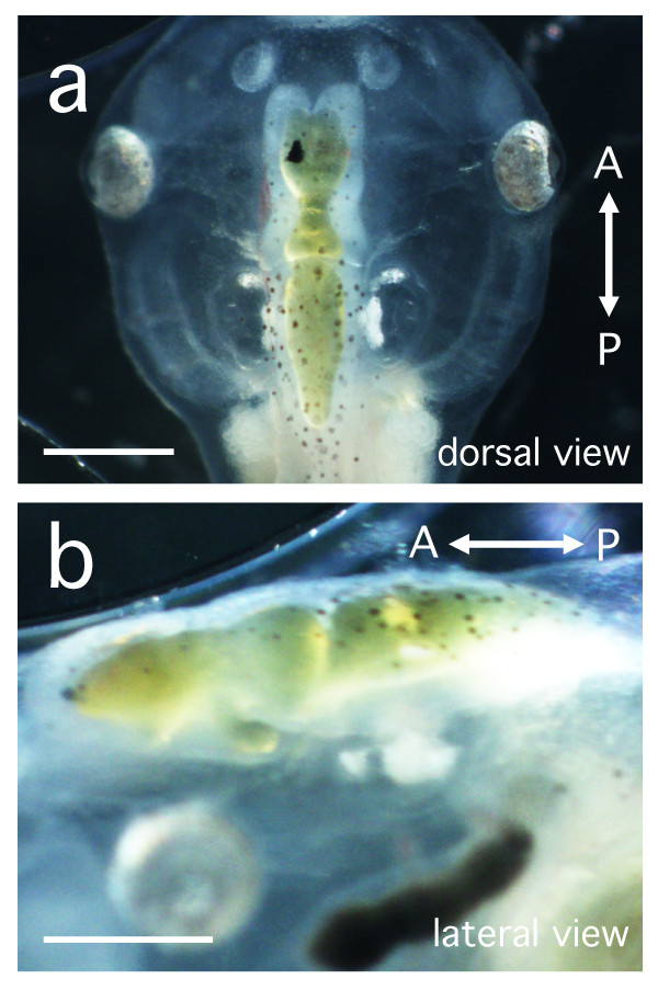
Background: It has long been known that cerebrospinal fluid (CSF), its composition and flow, play an important part in normal brain development, and ependymal cell ciliary beating as a possible driver of CSF flow has previously been studied in mammalian fetuses in vitro. Lower vertebrate animals are potential models for analysis of CSF flow during development because they are oviparous. Albino Xenopus laevis larvae are nearly transparent and have a straight, translucent brain that facilitates the observation of fluid flow within the ventricles. The aim of these experiments was to study CSF flow and circulation in vivo in the developing brain of living embryos, larvae and tadpoles of Xenopus laevis using a microinjection technique.
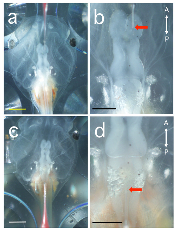
Methods: The development of Xenopus larval brain ventricles and the patterns of CSF flow were visualised after injection of quantum dot nanocrystals and polystyrene beads (3.1 or 5.8 μm in diameter) into the fourth cerebral ventricle at embryonic/larval stages 30-53.
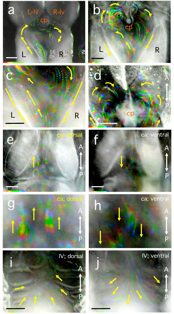
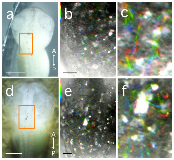
Results: The fluorescent nanocrystals showed the normal development of the cerebral ventricles from embryonic/larval stages 38 to 53. The polystyrene beads injected into stage 47-49 larvae revealed three CSF flow patterns, left-handed, right-handed and non-biased, in movement of the beads into the third ventricle from the cerebral aqueduct (aqueduct of Sylvius). In the lateral ventricles, anterior to the third ventricle, CSF flow moved anteriorly along the outer wall of the ventricle to the inner wall and then posteriorly, creating a semicircle. In the cerebral aqueduct, connecting the third and fourth cerebral ventricles, CSF flow moved rostrally in the dorsal region and caudally in the ventral region. Also in the fourth ventricle, clear dorso-ventral differences in fluid flow pattern were observed.
Conclusions: This is the first visualisation of the orchestrated CSF flow pattern in developing vertebrates using a live animal imaging approach. CSF flow in Xenopus albino larvae showed a largely consistent pattern, with the exception of individual differences in left-right asymmetrical flow in the third ventricle.
Figures

Similar articles
-
In Xenopus ependymal cilia drive embryonic CSF circulation and brain development independently of cardiac pulsatile forces.Fluids Barriers CNS. 2020 Dec 11;17(1):72. doi: 10.1186/s12987-020-00234-z. Fluids Barriers CNS. 2020. PMID: 33308296 Free PMC article.
-
Cerebrospinal fluid contacting neurons in the reduced brain ventricular system of the atlantic hagfish, Myxine glutinosa.Acta Biol Hung. 2003;54(1):35-44. doi: 10.1556/ABiol.54.2003.1.4. Acta Biol Hung. 2003. PMID: 12705320
-
Ciliogenesis and cerebrospinal fluid flow in the developing Xenopus brain are regulated by foxj1.Cilia. 2013 Sep 24;2(1):12. doi: 10.1186/2046-2530-2-12. Cilia. 2013. PMID: 24229449 Free PMC article.
-
Ependymal Cilia: Physiology and Role in Hydrocephalus.Front Mol Neurosci. 2022 Jul 12;15:927479. doi: 10.3389/fnmol.2022.927479. eCollection 2022. Front Mol Neurosci. 2022. PMID: 35903173 Free PMC article. Review.
-
The system of cerebrospinal fluid-contacting neurons. Its supposed role in the nonsynaptic signal transmission of the brain.Histol Histopathol. 2004 Apr;19(2):607-28. doi: 10.14670/HH-19.607. Histol Histopathol. 2004. PMID: 15024719 Review.
Cited by
-
Toxicity Effects of Functionalized Quantum Dots, Gold and Polystyrene Nanoparticles on Target Aquatic Biological Models: A Review.Molecules. 2017 Aug 31;22(9):1439. doi: 10.3390/molecules22091439. Molecules. 2017. PMID: 28858240 Free PMC article. Review.
-
Three-dimensional, three-vector-component velocimetry of cilia-driven fluid flow using correlation-based approaches in optical coherence tomography.Biomed Opt Express. 2015 Aug 24;6(9):3515-38. doi: 10.1364/BOE.6.003515. eCollection 2015 Sep 1. Biomed Opt Express. 2015. PMID: 26417520 Free PMC article.
-
Embryonic and skeletal development of the albino African clawed frog (Xenopus laevis).J Anat. 2023 Jun;242(6):1051-1066. doi: 10.1111/joa.13835. Epub 2023 Jan 28. J Anat. 2023. PMID: 36708289 Free PMC article.
-
Adeno-associated viral tools to trace neural development and connectivity across amphibians.Dev Cell. 2025 Mar 10;60(5):794-812.e6. doi: 10.1016/j.devcel.2024.10.025. Epub 2024 Nov 26. Dev Cell. 2025. PMID: 39603234
-
SCO-spondin from embryonic cerebrospinal fluid is required for neurogenesis during early brain development.Front Cell Neurosci. 2013 Jun 3;7:80. doi: 10.3389/fncel.2013.00080. eCollection 2013. Front Cell Neurosci. 2013. PMID: 23761733 Free PMC article.
References
-
- Taketomo T, Saito A. Experimental studies on cerebrospinal fluid flow. Neurology. 1965;15:578–586. - PubMed
-
- Bradley WG Jr, Scalzo D, Queralt J, Nitz WN, Atkinson DJ, Wong P. Normal-pressure hydrocephalus: evaluation with cerebrospinal fluid flow measurements at MR imaging. Radiology. 1996;198:523–529. - PubMed
LinkOut - more resources
Full Text Sources
