Review
. 2012 Apr 26;12(6):401-10.
doi: 10.1038/nrc3262.

Deconvoluting the context-dependent role for autophagy in cancer

Affiliations
Review

Deconvoluting the context-dependent role for autophagy in cancer

Eileen White. Nat Rev Cancer. .

Abstract

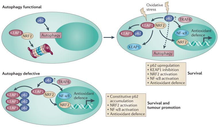
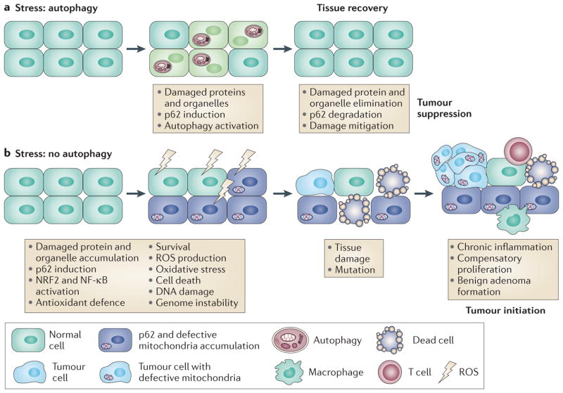
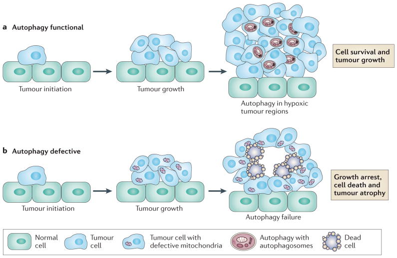
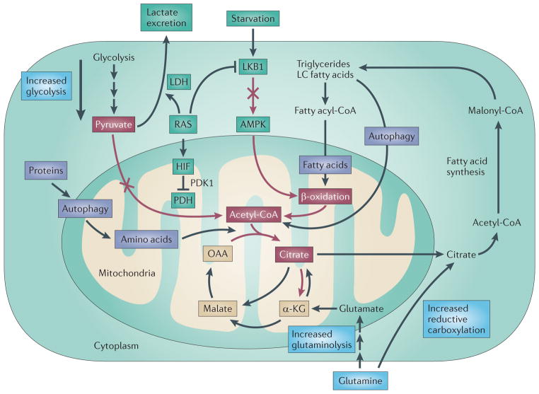
Autophagy (also known as macroautophagy) captures intracellular components in autophagosomes and delivers them to lysosomes, where they are degraded and recycled. Autophagy can have two functions in cancer. It can be tumour suppressive through the elimination of oncogenic protein substrates, toxic unfolded proteins and damaged organelles. Alternatively, it can be tumour promoting in established cancers through autophagy-mediated intracellular recycling that provides substrates for metabolism and that maintains the functional pool of mitochondria. Therefore, defining the context-specific role for autophagy in cancer and the mechanisms involved will be important to guide autophagy-based therapeutic intervention.


Conflict of interest statement

Competing interests statement

The author declares no competing financial interests.

Figures

Figure 1
Figure 1. Regulation of NRF2 by autophagy, KEAP1 and p62
Under normal conditions, nuclear factor erythroid 2-related factor 2 (NRF2) is bound to kelch-like ECH-associated protein 1 (KEAP1) and is inactivated as a transcription factor for antioxidant-defence genes by proteasome-mediated degradation. p62 is degraded through autophagy under normal conditions. In the presence of oxidative stress, KEAP1 is either modified so that it can no longer bind NRF2 or it is sequestered by p62, the expression of which is increased in response to oxidative stress. This displaces KEAP1 from NRF2 so that NRF2 can activate antioxidant-defence genes and promote survival. Oxidative stress also activates nuclear factor-κB (NF-κB) as a result of p62 upregulation and tumour necrosis factor receptor-associated factor 6 (TRAF6) complex formation, or by other mechanisms, to turn on antioxidant-defence gene expression. p62 is still subject to autophagy in cells experiencing cellular stress (dashed arrow). In autophagy-defective cells and tissues, the autophagy substrate p62 is not degraded, and so accumulates to high levels. p62 binds and sequesters KEAP1 in aggregates, resulting in the constitutive activation of NRF2 and antioxidant defence. This p62 induction can also activate NF-κB, antioxidant defence and survival.
Figure 2
Figure 2. Mechanism of autophagy-mediated tumour suppression
a| Autophagy, either basal or stress-induced, prevents the accumulation of oncogenic proteins such as p62, as well as damaged proteins and organelles. b | In autophagydefective tissues, p62 and damaged proteins and organelles accumulate. This is associated with the activation of oncogenic signalling pathways (nuclear factor erythroid 2-related factor 2 (NRF2) and nuclear factor-κB (NF-κB)) that promote survival but that are probably eventually overwhelmed by sustained oxidative stress. This leads to reactive oxygen species (ROS) production, chronic tissue damage, inflammation and genome instability, creating a tumour-initiating and tumour-promoting environment.
Figure 3
Figure 3. The role of autophagy in supporting the growth of aggressive cancers
a | Autophagy is upregulated in RAS-driven cancers and is also induced in hypoxic tumour regions where it supports tumour cell survival. b | Autophagydeficient tumour cells accumulate defective mitochondria and are prone to cell death in hypoxic regions. This can lead to impairment of the growth of RAS-driven cancers and perhaps other cancers.
Figure 4
Figure 4. Mechanism of autophagy addiction of RAS-driven cancers
The generation and use of acetyl CoA (depicted by red arrows and red boxes) is an essential component of the tricarboxylic acid (TCA) cycle. There are three known mechanisms by which RAS diminishes the pool of acetyl-CoA (shown in the green boxes). First, RAS can activate lactate dehydrogenase (LDH) that converts pyruvate to lactate, which is excreted. Second, RAS can activate hypoxia-inducible factor (HIF), inhibiting pyruvate dehydrogenase (PDH) and the conversion of pyruvate to acetyl-CoA. Third, RAS inhibits liver kinase B1 (LKB1), blocking AMP kinase (AMPK) and β-oxidation. Autophagy defects result in reduced citrate levels, impaired TCA cycle function and loss of mitochondrial respiration,. Autophagy can potentially compensate for the metabolic reprogramming by RAS by degrading proteins and lipids that provide amino acid and fatty acid substrates, producing acetyl-CoA (purple boxes). Tumour cells might also compensate for autophagy impairment by upregulating glycolysis, glutaminolysis or reductive carboxylation of α-ketoglutarate (α-KG) from glutamine (blue boxes). LC, long chain; OAA, oxaloacetate; PDK1, pyruvate dehydrogenase kinase 1.

References

    1. Levine B, Kroemer G. Autophagy in the pathogenesis of disease. Cell. 2008;132:27–42. - PMC - PubMed
    1. Mizushima N. The role of the Atg1/ULK1 complex in autophagy regulation. Curr Opin Cell Biol. 2010;22:132–139. - PubMed
    1. Rabinowitz JD, White E. Autophagy and metabolism. Science. 2010;330:1344–1348. - PMC - PubMed
    1. Mathew R, White E. Autophagy in tumorigenesis and energy metabolism: friend by day, foe by night. Curr Opin Genet Dev. 2011;21:113–119. - PMC - PubMed
    1. Clague MJ, Urbe S. Ubiquitin: same molecule, different degradation pathways. Cell. 2010;143:682–685. - PubMed

Publication types

LinkOut - more resources