. 2012 Aug;11(8):411-21.
doi: 10.1074/mcp.M111.013581. Epub 2012 Apr 24.

DNA ends alter the molecular composition and localization of Ku multicomponent complexes

Affiliations

DNA ends alter the molecular composition and localization of Ku multicomponent complexes

Guillaume Adelmant et al. Mol Cell Proteomics. 2012 Aug.

Abstract

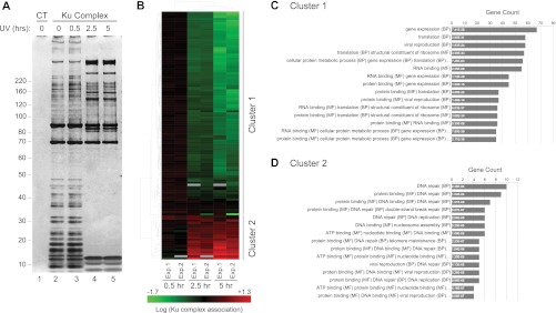
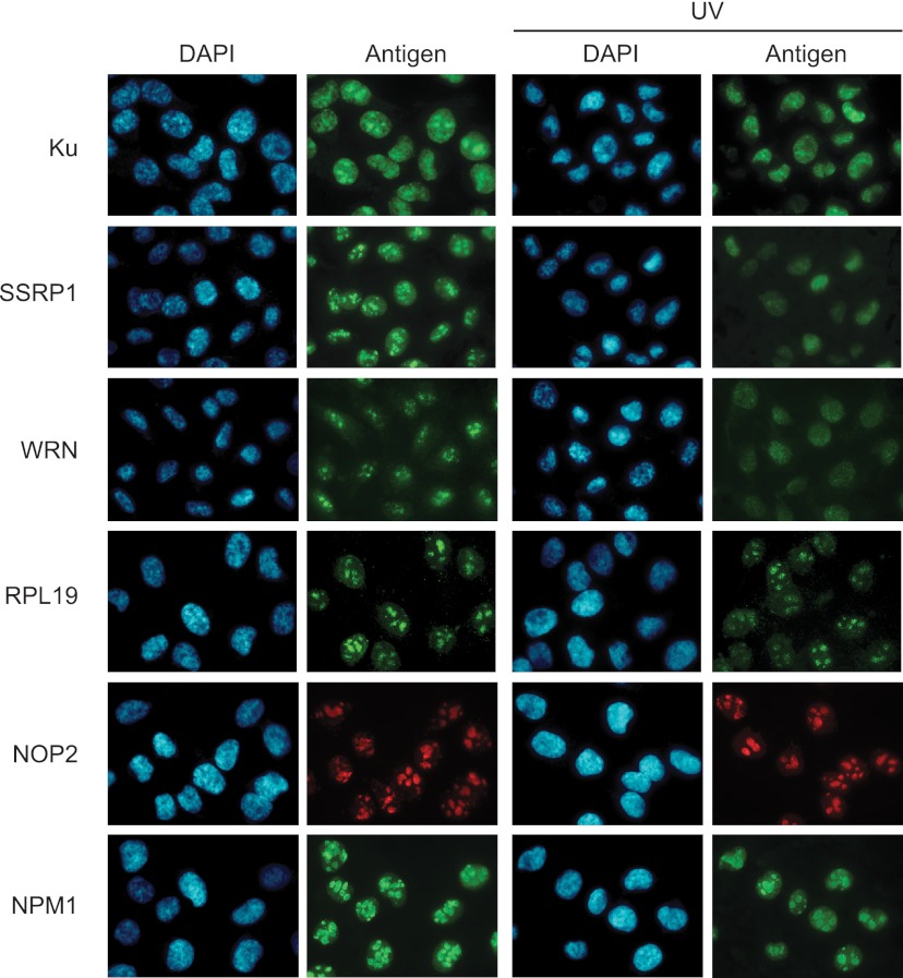
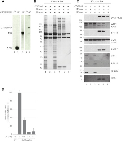
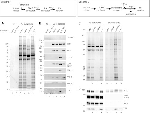
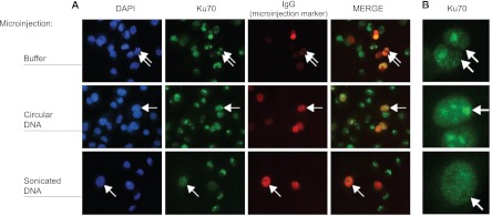
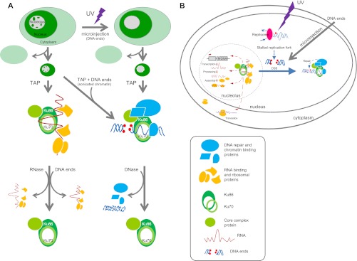
The Ku heterodimer plays an essential role in non-homologous end-joining and other cellular processes including transcription, telomere maintenance and apoptosis. While the function of Ku is regulated through its association with other proteins and nucleic acids, the specific composition of these macromolecular complexes and their dynamic response to endogenous and exogenous cellular stimuli are not well understood. Here we use quantitative proteomics to define the composition of Ku multicomponent complexes and demonstrate that they are dramatically altered in response to UV radiation. Subsequent biochemical assays revealed that the presence of DNA ends leads to the substitution of RNA-binding proteins with DNA and chromatin associated factors to create a macromolecular complex poised for DNA repair. We observed that dynamic remodeling of the Ku complex coincided with exit of Ku and other DNA repair proteins from the nucleolus. Microinjection of sheared DNA into live cells as a mimetic for double strand breaks confirmed these findings in vivo.


Figures

Fig. 1.
Fig. 1.
UV irradiation alters the composition of Ku86 macromolecular complexes. A, Protein complexes were purified from parental HeLa-S3 cells or HeLa-S3 cells expressing doubly tagged FLAG-HA Ku86 (supplemental Fig. S1). Representative silver stain analysis of nuclear Ku86 complexes purified before (t = 0, lane 2) or after (t = 0.5 h, 2.5 h, 5 h, lanes 3, 4, and 5, respectively) UV irradiation (300 J/m2), along with a “mock” complex purified from parental HeLa-S3 cells (lane 1). B, Hierarchical clustering of quantitative proteomic data obtained after iTRAQ LC-MS/MS analysis of biological replicates: 118 Ku86 associated proteins detected in two independent experiments (Exp.1 and Exp.2) primarily segregate between two clusters based on their response to UV irradiation. Green and red, respectively, indicate decreased and increased association after UV irradiation (see also supplemental Table S1 Summary of quantitative LC-MS data). (C and D) Functional enrichment analysis of proteins from Cluster 1 and 2. The number of genes represented in (B) for each category is provided on the horizontal axis. The corresponding adjusted p value is indicated within each bar. For clarity, only the first three co-occurrence terms are listed. See supplemental Table S2 Functional enrichment analysis, for the full list of annotations.
Fig. 2.
Fig. 2.
UV irradiation mediates nuclear re-localization of Ku complex components. Immunofluorescence analysis of Ku complex components in response to UV radiation. MDA-MB-231 cells were exposed to UV radiation and processed 2.5 h later for immunofluorescence following a triton extraction and paraformaldehyde fixation protocol (T/PFA). Ku, WRN and the DNA binding protein SSRP1 re-localize from the nucleolus to the nucleoplasm after irradiation with UV. The nucleolar and ribosomal proteins RPL19 and NOP2, which interact primarily with Ku in undamaged cells remain localized in the nucleolus after UV irradiation. The stability of staining for the nucleolar marker NPM1 indicates that nucleolar integrity is preserved after UV radiation.
Fig. 3.
Fig. 3.
Ku exists as distinct ribonucleo- and deoxyribonucleo-protein complexes. A, Northern blot analysis of RNA co-precipitating with Ku. Complexes were purified from nuclear extracts of parental HeLa-S3 (CT, lanes 1 and 4) or HeLa-S3 expressing Ku86-FLAG-HA cells (Ku) and Northern blot was used to probe for ribosomal RNA (5.8S and 18S, lanes 2 and 3) and U3snoRNA (lane 5). B, Silver stain analysis of nuclear Ku86 complexes purified before (lanes 1–3) or 5 h after UV irradiation (lanes 4–6). The complexes were incubated with buffer alone (lanes 1 and 4) or with RNase (lanes 2 and 5) or DNase (lanes 3 and 6) prior to tandem affinity purification. C, Western blot analysis of the Ku complexes shown in (B). Complexes were probed for DNA-PKcs, SPT16, SSRP1, WRN, RHA, Ku86-FLAG-HA, YB-1, RPL26, RPL19 and H2A. D, Quantitative RT-PCR analysis of 18S rRNA in nuclear Ku86 complexes after UV irradiation. Nuclear Ku complexes along with a “mock” complex were purified from parental HeLa-S3 cells as described in Fig. 1A. Copurifying 18S RNA was quantified after reverse transcription by real-time RT-PCR. Average abundance and standard deviation across three technical replicates is plotted relative to the normalized abundance of 18S present in the complex at t = 0.
Fig. 4.
Fig. 4.
DNA ends induce coordinate substitution of RNA associated proteins with DNA binding factors in Ku multicomponent complexes. (A and B: Scheme 1) (C and D: Scheme 2) Biochemical manipulation of nuclear extracts to monitor assembly of Ku complexes in the presence of intact or sonicated chromatin. A, Silver stain analysis of control (lanes 1 and 2) or nuclear Ku (lanes 3–6) complexes purified in the presence of buffer (lane 3) or intact (lanes 1 and 4), sonicated (lane 2 and 5) or UV irradiated (lane 6) chromatin. B, Western blot analysis of the protein complexes shown in (A). Complexes were probed for DNA-PKcs, SPT16, SSRP1, WRN, RHA, Ku86-FLAG-HA, RPL26, RPL19, and H2A. (C and D) Biochemical manipulation of immobilized Ku complexes to identify factors stably bound or released in response to incubation with DNA mimicking double-strand breaks. C, Silver stain analysis of complexes and supernatants resulting from incubation of immobilized complexes with buffer alone (lanes 1 and 5) or with circular (lanes 2 and 6), sonicated (lanes 3 and 7), or UV irradiated (lanes 4 and 8) plasmid DNA. D, Western blot analysis of the protein complexes and supernatants shown in (C).
Fig. 5.
Fig. 5.
DNA ends are sufficient to induce exit of Ku from the nucleolus in vivo. A, MDA-MB-231 cells were microinjected with rabbit immunoglobulin (IgG) as an injection marker, alone (top row) or in combination with circular (second row) or sonicated (third row) plasmid DNA and processed for immunofluorescence following a triton extraction and paraformaldehyde fixation protocol (T/PFA). Cells were labeled with an anti-rabbit IgG antibody to identify microinjected cells (red channel) and with an anti-mouse IgG antibody to detect endogenous Ku70 (green channel). Nuclei were stained with DAPI (blue channel). B, Cropped views of microinjected cells from (A) showing highly magnified nuclei. Arrows indicate representative microinjected cells.
Fig. 6.
Fig. 6.
Schematic summary of (A) experiments and observations in support of (B) the hypothesis that DNA ends alter the molecular composition and nuclear localization of Ku multicomponent complexes.

References

    1. Yano K., Morotomi-Yano K., Wang S. Y., Uematsu N., Lee K. J., Asaithamby A., Weterings E., Chen D. J. (2008) Ku recruits XLF to DNA double-strand breaks. EMBO Rep. 9, 91–96 - PMC - PubMed
    1. Calsou P., Delteil C., Frit P., Drouet J., Salles B. (2003) Coordinated assembly of Ku and p460 subunits of the DNA-dependent protein kinase on DNA ends is necessary for XRCC4-ligase IV recruitment. J. Mol. Biol. 326, 93–103 - PubMed
    1. Ma Y., Lu H., Tippin B., Goodman M. F., Shimazaki N., Koiwai O., Hsieh C. L., Schwarz K., Lieber M. R. (2004) A biochemically defined system for mammalian nonhomologous DNA end joining. Mol. Cell 16, 701–713 - PubMed
    1. Meek K., Gupta S., Ramsden D. A., Lees-Miller S. P. (2004) The DNA-dependent protein kinase: the director at the end. Immunol. Rev. 200, 132–141 - PubMed
    1. Chibazakura T., Watanabe F., Kitajima S., Tsukada K., Yasukochi Y., Teraoka H. (1997) Phosphorylation of human general transcription factors TATA-binding protein and transcription factor IIB by DNA-dependent protein kinase–synergistic stimulation of RNA polymerase II basal transcription in vitro. Eur. J. Biochem. 247, 1166–1173 - PubMed

Publication types

MeSH terms