. 2012 May 10;485(7397):207-12.
doi: 10.1038/nature11010.

Molecular mechanism of ATP binding and ion channel activation in P2X receptors

Affiliations

Molecular mechanism of ATP binding and ion channel activation in P2X receptors

Motoyuki Hattori et al. Nature. .

Abstract

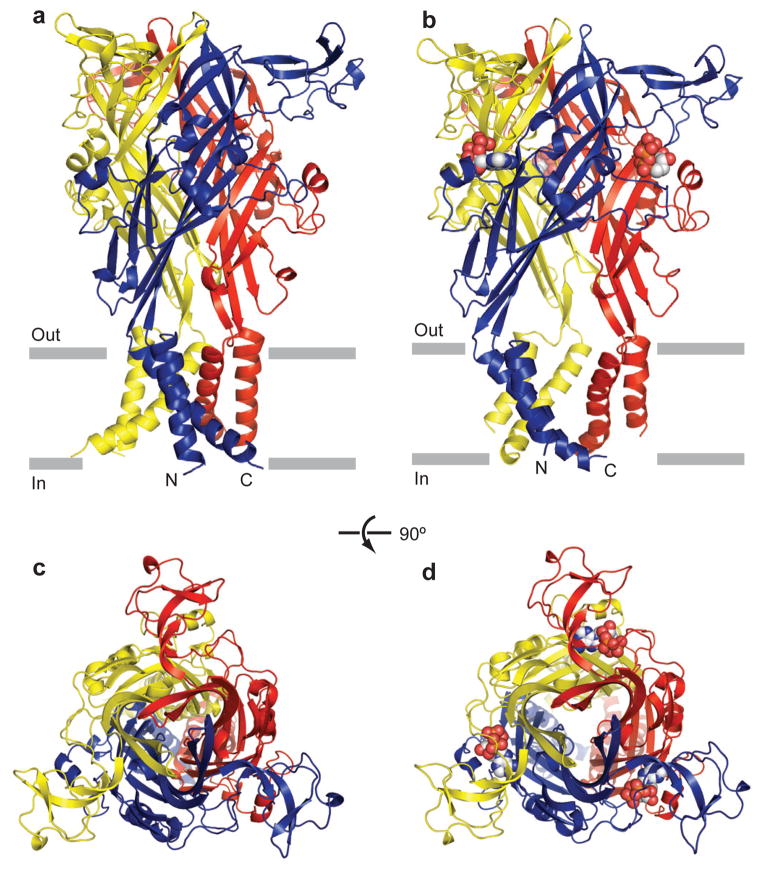
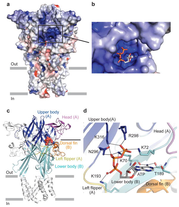
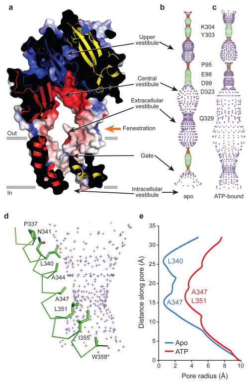
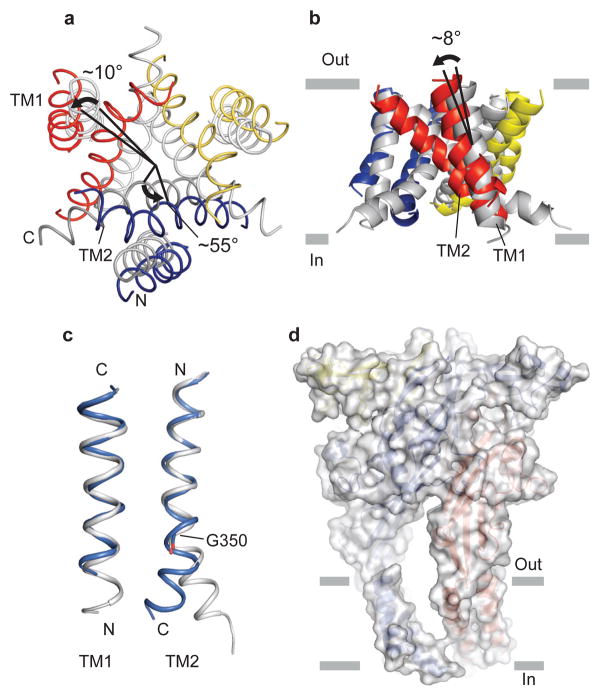
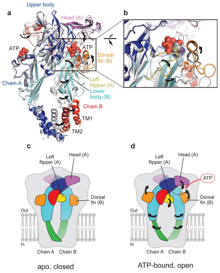
P2X receptors are trimeric ATP-activated ion channels permeable to Na+, K+ and Ca2+. The seven P2X receptor subtypes are implicated in physiological processes that include modulation of synaptic transmission, contraction of smooth muscle, secretion of chemical transmitters and regulation of immune responses. Despite the importance of P2X receptors in cellular physiology, the three-dimensional composition of the ATP-binding site, the structural mechanism of ATP-dependent ion channel gating and the architecture of the open ion channel pore are unknown. Here we report the crystal structure of the zebrafish P2X4 receptor in complex with ATP and a new structure of the apo receptor. The agonist-bound structure reveals a previously unseen ATP-binding motif and an open ion channel pore. ATP binding induces cleft closure of the nucleotide-binding pocket, flexing of the lower body β-sheet and a radial expansion of the extracellular vestibule. The structural widening of the extracellular vestibule is directly coupled to the opening of the ion channel pore by way of an iris-like expansion of the transmembrane helices. The structural delineation of the ATP-binding site and the ion channel pore, together with the conformational changes associated with ion channel gating, will stimulate development of new pharmacological agents.


Conflict of interest statement

The authors declare no competing financial interests

Figures

Figure 1
Figure 1. The architectures of zebrafish P2X4
a, b, zebrafish ΔP2X4-B2 (a) and ΔP2X4-C (b) trimer structures viewed parallel to the membrane. Each subunit is shown in a different color. ATP is shown in sphere representation. c, d, zebrafish ΔP2X4-B2(c) and ΔP2X4-C (d) trimer structures viewed from the extracellular side.
Figure 2
Figure 2. Analysis of conformational difference between the ΔP2X4-C and ΔP2X4-B2
Each structural feature of the dolphin-shaped P2X4 subunit is coloured differently. a, b, Superpositions between the ΔP2X4-C and ΔP2X4-B2 (grey) using Cα positions of the protomers (a) and trimers (b). c, d The TM and the body domains of ΔP2X4-B2 (c) and ΔP2X4-C (d). Only two subunits in the foreground are shown.
Figure 3
Figure 3. ATP binding site
a, b, An electrostatic potential surface of the ΔP2X4-C (a), contoured from −10 kT (red) to +10 kT (blue) (dielectric constant:80), and its close-up view (b). c, The regions forming the ATP-binding pocket are coloured as in Fig. 2a. The ATP molecule is shown in sphere representation. d, Close-up view of the ATP-binding site. The oxygen atom from the glycerol molecule is shown in sphere representation. Black dashed lines indicate hydrogen bonding (<3.3Å).
Figure 4
Figure 4. The transmembrane pore
a, A section of an electrostatic potential surface of the ΔP2X4-C, contoured as in Fig 3a. Pore-lining surfaces of ΔP2X4-B2 (b) and ΔP2X4-C (c). Each color indicates a different radius range from the pore center (red:<1.15Å, green: 1.15–2.3 Å, and purple:>2.3Å). d, Pore-lining residues of ΔP2X4-C shown in stick representation with the pore-lining surface. e, Pore radius for ΔP2X4-C and ΔP2X4-B2 along the pore center axis.
Figure 5
Figure 5. Structural transition in the TM domain
a, b, The TM region of ΔP2X4-C and ΔP2X4-B2 (grey), viewed from the intracellular side (a) and from parallel to the membrane (b). ΔP2X4-B2 is superimposed on ΔP2X4-C using Cα positions of the trimer. The black arrows and bars denote the rotation of the TM helices (a) and the orientation of the TM helices (b), respectively. c, Close-up view of the TM helices. TM helices of ΔP2X4-B2 (grey) are superimposed on those of ΔP2X4-C using Cα positions of residue 36–55 for TM1 and residue 334–349 for TM2, respectively. Gly350 is shown in stick representation. d, A surface model of the ΔP2X4-C trimer with the cartoon representation inside.
Figure 6
Figure 6. Mechanism of activation
a, A ΔP2X4-B2 subunit (grey) is superimposed on ΔP2X4-C using Cα positions of protomer A. Only two subunits in the foreground are shown. The rotation axis describes the superposition of the apo ΔP2X4-B2 B subunit onto the ATP-bound ΔP2X4-C B protomer. b, Close-up view of the conformational changes resulting from ATP binding. c, d A cartoon model of the ATP-dependent activation mechanism. The black arrows denote the movement from the apo closed state (c) to the ATP-bound open state (d).

References

    1. Burnstock G. Purinergic nerves. Pharmacol Rev. 1972;24:509–581. - PubMed
    1. Valera S, et al. A new class of ligand-gated ion channel defined by P2x receptor for extracellular ATP. Nature. 1994;371:516–519. - PubMed
    1. Webb TE, et al. Cloning and functional expression of a brain G-protein-coupled ATP receptor. FEBS Lett. 1993;324:219–225. - PubMed
    1. Surprenant A, North RA. Signaling at purinergic P2X receptors. Annu Rev Physiol. 2009;71:333–359. - PubMed
    1. Burnstock G, Kennedy C. P2X receptors in health and disease. Adv Pharmacol. 2011;61:333–372. - PubMed

Publication types