Characterization of extrastriatal D2 in vivo specific binding of [¹⁸F](N-methyl)benperidol using PET
- PMID: 22535514
- PMCID: PMC3389593
- DOI: 10.1002/syn.21566
Characterization of extrastriatal D2 in vivo specific binding of [¹⁸F](N-methyl)benperidol using PET
Abstract
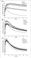
PET imaging studies of the role of the dopamine D2 receptor family in movement and neuropsychiatric disorders are limited by the use of radioligands that have near-equal affinities for D2 and D3 receptor subtypes and are susceptible to competition with endogenous dopamine. By contrast, the radioligand [¹⁸F]N-methylbenperidol ([¹⁸F]NMB) has high selectivity and affinity for the D2 receptor subtype (D2R) and is not sensitive to endogenous dopamine. Although [¹⁸F]NMB has high binding levels in striatum, its utility for measuring D2R in extrastriatal regions is unknown. A composite MR-PET image was constructed across 14 healthy adult participants representing average NMB uptake 60 to 120 min after [¹⁸F]NMB injection. Regional peak radioactivity was identified using a peak-finding algorithm. FreeSurfer and manual tracing identified a priori regions of interest (ROI) on each individual's MR image and tissue activity curves were extracted from coregistered PET images. [¹⁸F]NMB binding potentials (BP(ND) s) were calculated using the Logan graphical method with cerebellum as reference region. In eight unique participants, extrastriatal BP(ND) estimates were compared between Logan graphical methods and a three-compartment kinetic tracer model. Radioactivity and BP(ND) levels were highest in striatum, lower in extrastriatal subcortical regions, and lowest in cortical regions relative to cerebellum. Age negatively correlated with striatal BP(ND) s. BP(ND) estimates for extrastriatal ROIs were highly correlated across kinetic and graphical methods. Our findings indicate that PET with [¹⁸F]NMB measures specific binding in extrastriatal regions, making it a viable radioligand to study extrastriatal D2R levels in healthy and diseased states.
Copyright © 2012 Wiley Periodicals, Inc.
Figures


References
-
- Antonini A, Leenders KL. Dopamine D2 receptors in normal human brain: Effect of age measured by positron emission tomography (PET) and [11C]-raclopride. Ann NY Acad Sci. 1993;695:81–85. - PubMed
-
- Antonini A, Leenders KL, Reist H, Thomann R, Beer HF, Locher J. Effect of age on D2 dopamine receptors in normal human brain measured by positron emission tomography and 11C-raclopride. Arch Neurol. 1993;50:474–480. - PubMed
-
- Arnett CD, Shiue CY, Wolf AP, Fowler JS, Logan J, Watanabe M. Comparison of three 18F-labeled butyrophenone neuroleptic drugs in the baboon using positron emission tomography. J Neurochem. 1985;44:835–844. - PubMed
-
- Arnett CD, Wolf AP, Shiue CY, Fowler JS, MacGregor RR, Christman DR, Smith MR. Improved delineation of human dopamine receptors using [18F]-N-methylspiroperidol and PET. J Nucl Med. 1986;27:1878–1882. - PubMed
Publication types
MeSH terms
Substances
Grants and funding
- K24 MH087913/MH/NIMH NIH HHS/United States
- R01 DK085575/DK/NIDDK NIH HHS/United States
- T32 DA007261/DA/NIDA NIH HHS/United States
- R01 NS031001/NS/NINDS NIH HHS/United States
- R01 NS058714/NS/NINDS NIH HHS/United States
- 5 K24 MH087913-02/MH/NIMH NIH HHS/United States
- R01NS41509/NS/NINDS NIH HHS/United States
- R01DK085575/DK/NIDDK NIH HHS/United States
- R01 NS075321/NS/NINDS NIH HHS/United States
- R01NS075321/NS/NINDS NIH HHS/United States
- R01NS058714/NS/NINDS NIH HHS/United States
- R01 NS041509/NS/NINDS NIH HHS/United States
- R01NS031001/NS/NINDS NIH HHS/United States
- P30 NS048056/NS/NINDS NIH HHS/United States
- 5 T32 DA 007261-20/DA/NIDA NIH HHS/United States
LinkOut - more resources
Full Text Sources
