Review
. 2012 Jul;93(Pt 7):1385-1409.
doi: 10.1099/vir.0.042499-0. Epub 2012 Apr 25.

Non-canonical translation in RNA viruses

Affiliations
Review

Non-canonical translation in RNA viruses

Andrew E Firth et al. J Gen Virol. 2012 Jul.

Abstract

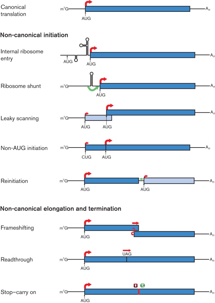
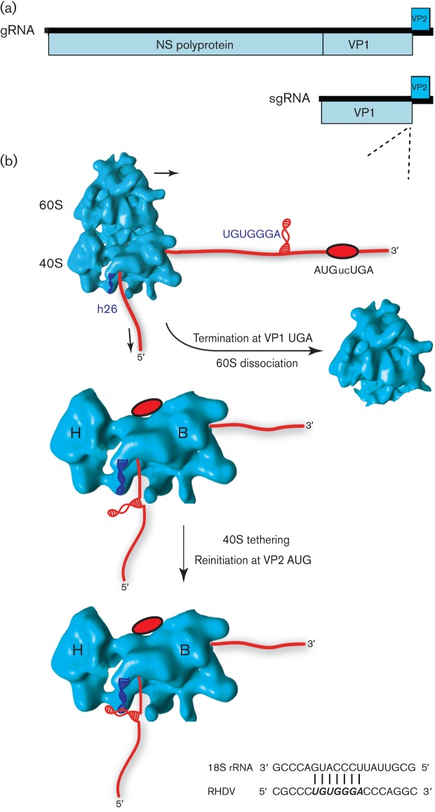
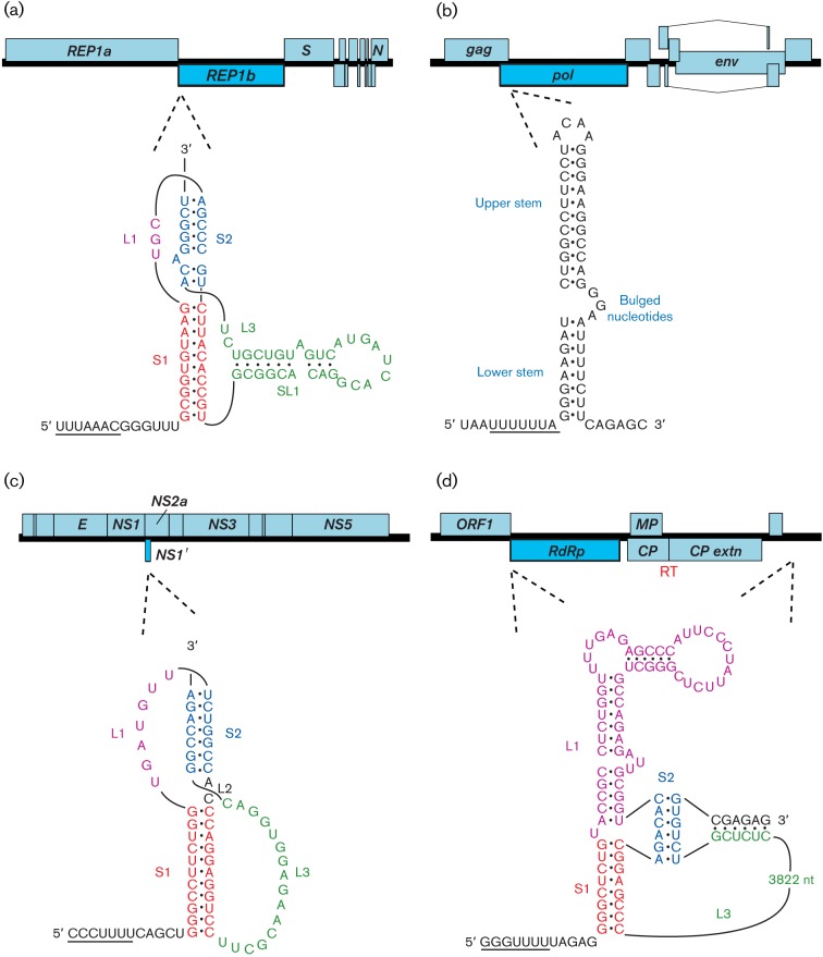
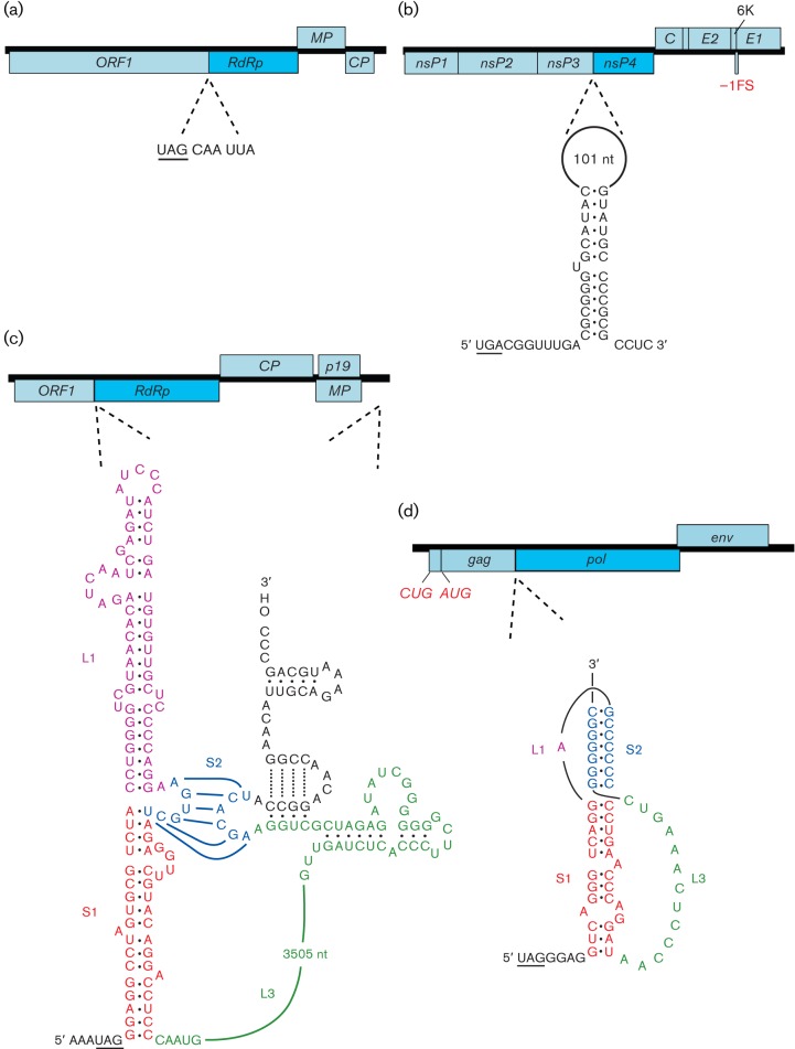
Viral protein synthesis is completely dependent upon the translational machinery of the host cell. However, many RNA virus transcripts have marked structural differences from cellular mRNAs that preclude canonical translation initiation, such as the absence of a 5' cap structure or the presence of highly structured 5'UTRs containing replication and/or packaging signals. Furthermore, whilst the great majority of cellular mRNAs are apparently monocistronic, RNA viruses must often express multiple proteins from their mRNAs. In addition, RNA viruses have very compact genomes and are under intense selective pressure to optimize usage of the available sequence space. Together, these features have driven the evolution of a plethora of non-canonical translational mechanisms in RNA viruses that help them to meet these challenges. Here, we review the mechanisms utilized by RNA viruses of eukaryotes, focusing on internal ribosome entry, leaky scanning, non-AUG initiation, ribosome shunting, reinitiation, ribosomal frameshifting and stop-codon readthrough. The review will highlight recently discovered examples of unusual translational strategies, besides revisiting some classical cases.


Figures

Fig. 1.
Fig. 1.
Examples of non-canonical translational mechanisms utilized by RNA viruses. Canonical eukaryotic mRNA translation is shown in the top panel. Red arrows indicate initiation of protein synthesis (at the start of an ORF) or continuation of translation by 80S ribosomes, with thicker arrows indicating the predominant path taken by ribosomes (not to scale). Green arrows indicate the probable movement of 40S subunits in a non-canonical manner. Where two distinct polypeptides are synthesized, the ORFs are shown in different shades of blue; where a recoding event during elongation leads to C-terminal extension of a polypeptide, the two ORFs are shown in the same colour. In the stop–carry on mechanism, both termination and initiation steps are non-canonical, as indicated by the red square and green circle.
Fig. 2.
Fig. 2.
The Israeli acute paralysis dicistrovirus IGR-IRES directs translation of two overlapping ORFs. (a) Genome map. Distinct IRESes direct translation of non-structural and structural polyproteins. The IGR-IRES also directs translation of ORFx, which overlaps ORF2 in the +1 reading frame. (b) Schematic of the IGR-IRES, showing pseudoknots (PK) I, II and III and stem–loops (SL) III, IV, V and VI. PK I occupies the ribosomal P-site and translation of ORF2 initiates at the GGC codon in the ribosomal A-site. (c) The formation of an additional base pair in PK I (U-G; bold) leads instead to initiation at the +1 frame GCG codon and translation of ORFx. Modified from a figure kindly provided by E. Jan (Ren et al., 2012).
Fig. 3.
Fig. 3.
Genome map of Panicum mosaic panicovirus. MP1, MP2, CP and p15 are all expressed from a single sgRNA via a combination of leaky scanning and non-AUG initiation. Initiation codons are indicated in upper case and nucleotides that differ from a strong initiation context are indicated in red.
Fig. 4.
Fig. 4.
Ribosome shunting, reinitiation and leaky scanning in members of the family Caulimoviridae. (a) Translation of ORF I of the pgRNA of rice tungro bacilliform tungrovirus is by ribosome shunting. Here, 40S complexes released after translation of the short 5′-most ORF (yellow) are able to shunt past a stable stem–loop in the 5′UTR (red arrow) and continue scanning the mRNA. Reinitiation subsequently takes place at the start codons of either ORF I (a non-AUG codon, AUU), ORF II (weak context AUG) or ORF III (strong context AUG). ORF IV is expressed from a spliced mRNA. (b) In cauliflower mosaic caulimovirus, a similar shunting mechanism is used to access the 5′-most coding ORF, VII. However, downstream ORFs I–V are translated via reinitiation events that are stimulated by a viral reinitiation factor, transactivator viroplasmin (TAV), expressed from an sgRNA (see text).
Fig. 5.
Fig. 5.
Proposed model for termination–reinitiation in caliciviruses. (a) Genome map of the calicivirus rabbit hemorrhagic disease lagovirus (RHDV). Expression of VP2 is by termination–reinitiation during translation of the viral sgRNA. (b) As the ribosome approaches the termination–reinitiation site (red oval; AUGucUGA in RHDV), the stretch of RNA containing TURBS motif 1 (UGUGGGA), predicted to be located in an RNA secondary structure, is translated and may be remodelled. During termination, the secondary structure is located in the mRNA exit channel of the ribosome [located between the head (H) and body (B) of the 40S subunit] and in close proximity to the solvent-accessible helix 26 (h26) of 18S rRNA (indicated as a blue helix). Base pairing between complementary residues in motif 1 and h26 occurs (shown at the bottom), with the interaction likely to be stabilized by eIF3 (not shown), also known to contact the TURBS and 18S rRNA. Together, these interactions act to tether the ribosome to the viral RNA, preventing its dissociation, allowing time for the recruitment of initiation factors and subsequent reinitiation on the downstream VP2 ORF.
Fig. 6.
Fig. 6.
Programmed ribosomal frameshifting in viral mRNAs. In each of the four examples, the genome is indicated as an ORF map, with the location of the frameshift site shown by dotted lines. In SARS coronavirus (a), Japanese encephalitis flavivirus (c) and barley yellow dwarf luteovirus (d), frameshifting is stimulated by an RNA pseudoknot (including a long-range interaction in the luteovirus). In HIV-1 (b), frameshifting is stimulated by a two-stem helix, although the upper stem makes the major contribution to frameshifting efficiency. In each case, the slippery shift site sequence is underlined. Note that, in (c), the unprocessed frameshift product generates a truncated polyprotein, unlike the other examples, where the frameshift facilitates extension of the polyprotein. Spliced and subgenomic RNAs are not shown and polyprotein cleavage products are only indicated where specifically relevant. ‘RT’ indicates a stop codon readthrough site.
Fig. 7.
Fig. 7.
Programmed stop codon readthrough in viral mRNAs. In each of the four examples, the genome is indicated as an ORF map, with the location of the readthrough site shown by dotted lines. In tobacco mosaic tobamovirus (a), only a short local sequence context 3′ of the recoded UAG is required for efficient readthrough. In Venezuelan equine encephalitis alphavirus (b), carnation italian ringspot tombusvirus (c) and murine leukemia gammaretrovirus (d), the 3′ stimulator is an RNA secondary structure: an extended stem–loop in (b), an RNA pseudoknot in (d) and long-range base pairing in (c). Spliced and subgenomic RNAs are not shown and polyprotein cleavage products are only indicated where specifically relevant. ‘FS’ indicates a ribosomal frameshift site.

References

    1. Adams M. J., Antoniw J. F., Mullins J. G. (2001). Plant virus transmission by plasmodiophorid fungi is associated with distinctive transmembrane regions of virus-encoded proteins. Arch Virol 146, 1139–1153 10.1007/s007050170111 - DOI - PubMed
    1. Agranovsky A. A., Koonin E. V., Boyko V. P., Maiss E., Frötschl R., Lunina N. A., Atabekov J. G. (1994). Beet yellows closterovirus: complete genome structure and identification of a leader papain-like thiol protease. Virology 198, 311–324 10.1006/viro.1994.1034 - DOI - PubMed
    1. Ahlquist P. (2006). Parallels among positive-strand RNA viruses, reverse-transcribing viruses and double-stranded RNA viruses. Nat Rev Microbiol 4, 371–382 10.1038/nrmicro1389 - DOI - PMC - PubMed
    1. Ahmadian G., Randhawa J. S., Easton A. J. (2000). Expression of the ORF-2 protein of the human respiratory syncytial virus M2 gene is initiated by a ribosomal termination-dependent reinitiation mechanism. EMBO J 19, 2681–2689 10.1093/emboj/19.11.2681 - DOI - PMC - PubMed
    1. Alam S. L., Wills N. M., Ingram J. A., Atkins J. F., Gesteland R. F. (1999). Structural studies of the RNA pseudoknot required for readthrough of the gag-termination codon of murine leukemia virus. J Mol Biol 288, 837–852 10.1006/jmbi.1999.2713 - DOI - PubMed

Publication types

LinkOut - more resources