Review
. 2012 Apr;92(2):635-88.
doi: 10.1152/physrev.00008.2011.

Matricellular proteins in cardiac adaptation and disease

Affiliations
Review

Matricellular proteins in cardiac adaptation and disease

Nikolaos G Frangogiannis. Physiol Rev. 2012 Apr.

Abstract

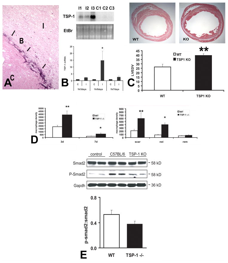
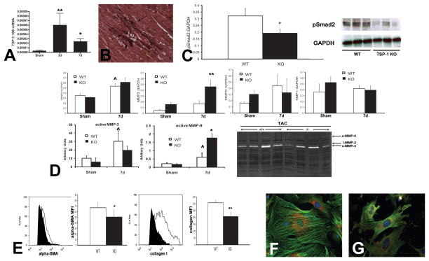
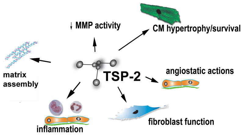
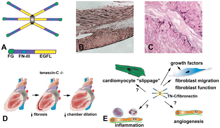
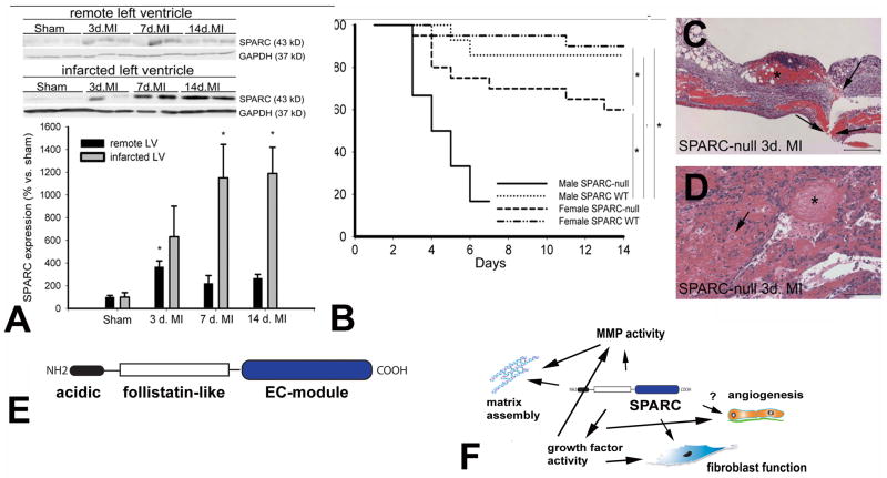
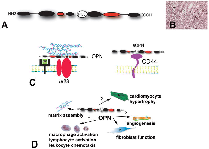
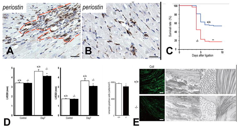
The term matricellular proteins describes a family of structurally unrelated extracellular macromolecules that, unlike structural matrix proteins, do not play a primary role in tissue architecture, but are induced following injury and modulate cell-cell and cell-matrix interactions. When released to the matrix, matricellular proteins associate with growth factors, cytokines, and other bioactive effectors and bind to cell surface receptors transducing signaling cascades. Matricellular proteins are upregulated in the injured and remodeling heart and play an important role in regulation of inflammatory, reparative, fibrotic and angiogenic pathways. Thrombospondin (TSP)-1, -2, and -4 as well as tenascin-C and -X secreted protein acidic and rich in cysteine (SPARC), osteopontin, periostin, and members of the CCN family (including CCN1 and CCN2/connective tissue growth factor) are involved in a variety of cardiac pathophysiological conditions, including myocardial infarction, cardiac hypertrophy and fibrosis, aging-associated myocardial remodeling, myocarditis, diabetic cardiomyopathy, and valvular disease. This review discusses the properties and characteristics of the matricellular proteins and presents our current knowledge on their role in cardiac adaptation and disease. Understanding the role of matricellular proteins in myocardial pathophysiology and identification of the functional domains responsible for their actions may lead to design of peptides with therapeutic potential for patients with heart disease.


Figures

Figure 1
Figure 1. Morphology of the normal mammalian heart
The adult heart contains cardiomyocytes, non-cardiomyocytes and a complex network of extracellular matrix. Each myocyte is surrounded by collagen (endomysium); individual fibers are also enmeshed in connective tissue (perimysium). Interactions between the matrix and cardiomyocytes are essential for their survival and function. Non-myocytes outnumber cardiomyocytes in the normal adult heart. The heart contains a rich vascular network comprised of capillary (c), venous and arteriolar endothelial cells, pericytes (P) and smooth muscle cells. A large number of resident fibroblasts is also noted (F). Normal mammalian hearts also contain small numbers of macrophages, mast cells (MC), lymphocytes and dendritic cells.
Figure 2
Figure 2. Structure of the TSPs
On the basis of their oligomerization status and architecture, TSPs are divided into trimeric (Group A) and pentameric (Group B) TSPs (see text). Abbreviations: NTD, N-terminal domain; vWF-C von Willebrand Factor homology domain; CTD, C-terminal domain.
Figure 3
Figure 3. The concept of “de-adhesion” in tissue remodeling
In remodeling tissues, induction of the prototypical matricellular proteins (TSP-1, tenascin-C, SPARC) may stimulate disassembly of focal adhesions and stress fibers in strongly adherent cells, inducing a state of intermediate cell adhesion. This process, called “de-adhesion” and may be important in promoting cell motility while preventing cell anoikis.
Figure 4
Figure 4. Main in vivo actions of TSP-1
A. TSP-1 is an important activator of TGF-β. After proteolytic cleavage of TGF-β from its propeptide, the TGF-β dimer remains bound to the Latency-Associated Peptide (LAP) by non-covalent interactions forming the small latent complex. TSP-1 binds to the sequence LSKL in the LAP and alters the conformation of TGF-β making it accessible to its receptors, TβRII and TβRI. Other TSPs do not exert TGF-β-activating effects. B. TSP-1 is a potent angiostatic agent through actions involving CD36. TSP-1 inhibits angiogenesis by modulating angiogenic growth factor (GF) signaling and by inducing endothelial cell apoptosis through a CD36/fyn/p38 mediated cascade. C. TSP-1 inhibits protease activity. TSP-1 inhibits MMP-3-dependent MMP-9 activation and attenuates thrombin-induced MMP-2 activation.
Figure 5
Figure 5. The role of TSP-1 in myocardial infarction
In the infarcted heart selective upregulation of TSP-1 in the infarct border zone may prevent expansion of the inflammatory infiltrate into the non-infarcted area A. Immunohistochemical staining of the infarcted canine heart demonstrates selective incorporation of TSP-1 (arrows) into the matrix of the infarct border zone (B). C, control non-infarcted myocardium; I, infarct B. Northern blotting shows marked TSP-1 upregulation in the infarcted canine myocardium. C. TSP-1 −/− mice exhibited accentuated dilative remodeling following myocardial infarction. D. Adverse remodeling in TSP-1 −/− mice was associated with expansion of the inflammatory infiltrate into the non-infarcted myocardium indicating failure of the protective “barrier” mechanism preventing expansion of the inflammatory infiltrate into the non-infarcted area. E. TSP-1 absence was associated with decreased Smad2 phosphorylation in the infarcted heart, suggesting impaired TGF-β signaling. TSP-1 deposition in the infarct border zone may protect the infarcted myocardium by inhibiting MMP activity, by exerting direct anti-inflammatory actions, by locally activating TGF-β (thus reducing macrophage inflammatory activity) or through inhibition of uncontrolled angiogenesis. The TSP-1 “barrier” may be responsible for containment of the inflammatory and angiogenic response within the infarct, thus preventing expansion of granulation tissue formation in the viable myocardium (Data reproduced with permission from Frangogiannis NG, Ren G, Dewald O, Zymek P, Haudek S, Koerting A, Winkelmann K, Michael LH, Lawler J, Entman ML. Critical role of endogenous thrombospondin-1 in preventing expansion of healing myocardial infarcts Circulation 2005;111:2935–42. Copyright 2005, American Heart Association).
Figure 6
Figure 6. The role of TSP-1 in cardiac fibrosis due to pressure overload
TSP-1 protects the pressure-overloaded myocardium by modulating fibroblast phenotype and matrix metabolism. A. qPCR shows marked TSP-1 upregulation in the pressure-overloaded myocardium in a mouse model of transverse aortic constriction. B. TSP-1 in the pressure-overloaded myocardium is localized in the cardiac interstitium. C. TSP-1 null mice exhibit worse dilative remodeling of the pressure-overloaded myocardium. Increased chamber dilation is associated with impaired TGF-β signaling (evidenced by reduced Smad2 phosphorylation). D. TSP-1 null animals exhibit increased MMP-9 activity in the pressure overloaded heart associated with accentuated MMP-3 levels. EG. Cardiac fibroblasts isolated from TSP-1 null pressure overloaded hearts are functionally impaired exhibiting reduced collagen expression and defective myofibroblast transdifferentiation. TSP-1 protects the pressure overloaded heart from chamber dilation by promoting TGF-β-induced myofibroblast transdifferentiation and activation and by inhibiting MMP activity. (Data reproduced with permission from Xia Y, Dobaczewski M, Gonzalez-Quesada C, Chen W, Biernacka A, Li, N, Lee DW, Frangogiannis NG. Endogenous thrombospondin-1 protects the pressure-overloaded myocardium by modulating fibroblast phenotype and matrix metabolism. Hypertension 2011;58: 902–911: Copyright 2011, American Heart Association)
Figure 7
Figure 7. Actions of TSP-2 in the remodeling myocardium
Experimental evidence using loss-of-function approaches suggests an important role for TSP-2 in protection of the aging, infarcted and pressure-overloaded heart. TSP-2 null mice develop dilative cardiomyopathy; this may be due to loss of CD47/integrin-mediated pro-survival signals in cardiomyocytes. TSP-2 absence is also associated with cardiac rupture and heart failure in models of myocardial infarction and angiotensin-II-mediated hypertrophy. TSP-2 may protect the remodeling heart by mediated essential actions on assembly and organization of the cardiac matrix, by inhibiting MMP activity, by activating pro-survival signals on cardiomyocytes, or by suppressing inflammation.
Figure 8
Figure 8. The role of tenascin-C in cardiac remodeling
A. tenascin-C assembles into a hexamer; each subunit contains EGF-like repeats (EGFL), a series of fibronectin type III modules (FN-III) and a C-terninal globular fibrinogen-like region (FG). B. Immunohistochemical staining for tenascin-C in reperfused mouse myocardial infarction illustrates that tenascin-C is selectively localized in the infarct border zone. C. In patients with ischemic cardiomyopathy, interstitial tenascin-C expression marks areas exhibiting active remodeling. D. Tenascin-C absence is associated with reduced fibrosis and attenuated chamber dilation following myocardial infarction. E. The effects of tenascin-C on the remodeling heart appear to be related to its profibrotic actions. Furthermore, tenascin-C may modulate the inflammatory and angiogenic response and may facilitate cardiomyocyte slippage.
Figure 9
Figure 9. The role of SPARC in cardiac remodeling
A. SPARC upregulation in the infarcted mouse myocardium. B-D. SPARC null mice have increased incidence of cardiac rupture (C, arrow), exhibiting intramural hemorrhages in the infarcted myocardium (D, arrows). Data reproduced with permission from: Schellings MW, Vanhoutte D, Swinnen M, Cleutjens JP, Debets J, van Leeuwen RE, d’Hooge J, Van de Werf F, Carmeliet P, Pinto YM, Sage EH, Heymans S. Absence of SPARC results in increased cardiac rupture and dysfunction after acute myocardial infarction. J Exp Med 2009;206:113–123: Copyright 2009, Rockefeller University Press. E. The SPARC molecule contains an acidic region, a follistatin-like domain and an extracellular Ca2+-binding region (EC-module). F. The mechanisms involved in SPARC-mediated cardiac fibrosis are likely due to enhanced growth factor signaling and to effects on matrix assembly, MMP activity and fibroblast function.
Figure 10
Figure 10. The role of OPN in cardiac remodeling
A. Structure of OPN. The RGD sequence is involved in several integrin-mediated actions of the OPN molecule. Ca2+-binding domains are indicated in red. B. Immunohistochemical staining for OPN in the infarcted canine myocardium. In the healing infarct, OPN is predominantly localized in macrophages and may be an indicator of their maturation and differentiation. C. OPN acts a) as a matricellular protein that binds to the matrix and modulates growth factor signaling and integrin-mediated actions and b) as a cytokine (soluble OPN) that signals through CD44. D. The role of OPN in cardiac remodeling has been investigated using loss-of-function models. OPN absence is associated with impaired formation of the collagenous scar following myocardial infarction leading to accentuated dilative remodeling. In models of cardiac pressure overload, OPN null animals exhibit attenuation of fibrosis and hypertrophy. Thus, the effects of OPN in cardiac remodeling appear to be mediated primarily through actions on matrix organization, fibroblast function and cardiomyocyte hypertrophy. The contribution of myocardial OPN upregulation in modulating the inflammatory and angiogenic response following cardiac injury remains unknown.
Figure 11
Figure 11. Role of periostin in myocardial infarction
A–B. Periostin expression if upregulated in the infarcted myocardium and is primarily localized in the infarct border zone (A) and in the remodeling myocardium (B). The dashed red line shows the infarct border zone. C. Periostin null mice exhibit a high incidence of cardiac rupture. D. However, surviving periostin −/− mice had attenuated dilative post-infarction remodeling. E. Reduced remodeling in periostin null mice was associated with attenuated fibroblast infiltration and smaller and less abundant collagen fibers in the infarct border zone. Data reproduced Data reproduced with permission from: Shimazaki M, Nakamura K, Kii I, Kashima T, Amizuka N, Li M, Saito M, Fukuda K, Nishiyama T, Kitajima S, Saga Y, Fukayama M, Sata M, Kudo A. Periostin is essential for cardiac healing after acute myocardial infarction. J Exp Med 2008;205:295:303: Copyright 2008, Rockefeller University Press.

References

    1. Abe K, Hibino T, Mishima H, Shimomura Y. The cytokine regulation of SPARC production by rabbit corneal epithelial cells and fibroblasts in vitro. Cornea. 2004;23:172–179. - PubMed
    1. Abel ED, Litwin SE, Sweeney G. Cardiac remodeling in obesity. Physiol Rev. 2008;88:389–419. - PMC - PubMed
    1. Abraham D. Connective tissue growth factor: growth factor, matricellular organizer, fibrotic biomarker or molecular target for anti-fibrotic therapy in SSc? Rheumatology (Oxford) 2008;47 (Suppl 5):v8–9. - PubMed
    1. Abreu JG, Ketpura NI, Reversade B, De Robertis EM. Connective-tissue growth factor (CTGF) modulates cell signalling by BMP and TGF-beta. Nat Cell Biol. 2002;4:599–604. - PMC - PubMed
    1. Adams JC. Thrombospondins: multifunctional regulators of cell interactions. Annu Rev Cell Dev Biol. 2001;17:25–51. - PubMed

Publication types

MeSH terms