

Classic and new animal models of Parkinson's disease
- PMID: 22536024
- PMCID: PMC3321500
- DOI: 10.1155/2012/845618
Classic and new animal models of Parkinson's disease
Abstract
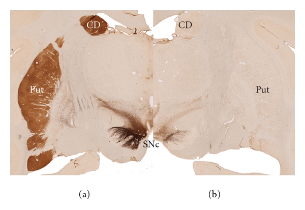
Neurological disorders can be modeled in animals so as to recreate specific pathogenic events and behavioral outcomes. Parkinson's Disease (PD) is the second most common neurodegenerative disease of an aging population, and although there have been several significant findings about the PD disease process, much of this process still remains a mystery. Breakthroughs in the last two decades using animal models have offered insights into the understanding of the PD disease process, its etiology, pathology, and molecular mechanisms. Furthermore, while cellular models have helped to identify specific events, animal models, both toxic and genetic, have replicated almost all of the hallmarks of PD and are useful for testing new neuroprotective or neurorestorative strategies. Moreover, significant advances in the modeling of additional PD features have come to light in both classic and newer models. In this review, we try to provide an updated summary of the main characteristics of these models as well as the strengths and weaknesses of what we believe to be the most popular PD animal models. These models include those produced by 6-hydroxydopamine (6-OHDA), 1-methyl-1,2,3,6-tetrahydropiridine (MPTP), rotenone, and paraquat, as well as several genetic models like those related to alpha-synuclein, PINK1, Parkin and LRRK2 alterations.
Figures
References
-
- Lees AJ, Hardy J, Revesz T. Parkinson’s disease. The Lancet. 2009;373(9680):2055–2066. - PubMed
-
- Spillantini MG, Schmidt ML, Lee VMY, Trojanowski JQ, Jakes R, Goedert M. α-synuclein in Lewy bodies [8] Nature. 1997;388(6645):839–840. - PubMed
-
- Forno LS. Neuropathology of Parkinson’s disease. Journal of Neuropathology and Experimental Neurology. 1996;55(3):259–272. - PubMed
-
- Braak H, Ghebremedhin E, Rüb U, Bratzke H, Del Tredici K. Stages in the development of Parkinson’s disease-related pathology. Cell and Tissue Research. 2004;318(1):121–134. - PubMed
-
- Chaudhuri KR, Healy DG, Schapira AHV. Non-motor symptoms of Parkinson’s disease: diagnosis and management. Lancet Neurology. 2006;5(3):235–245. - PubMed
Publication types
MeSH terms
Grants and funding
LinkOut - more resources
Full Text Sources
Other Literature Sources
Medical
