

Cone dysfunctions in retinitis pigmentosa with retinal nerve fiber layer thickening
- PMID: 22536039
- PMCID: PMC3334214
- DOI: 10.2147/OPTH.S28938
Cone dysfunctions in retinitis pigmentosa with retinal nerve fiber layer thickening
Abstract
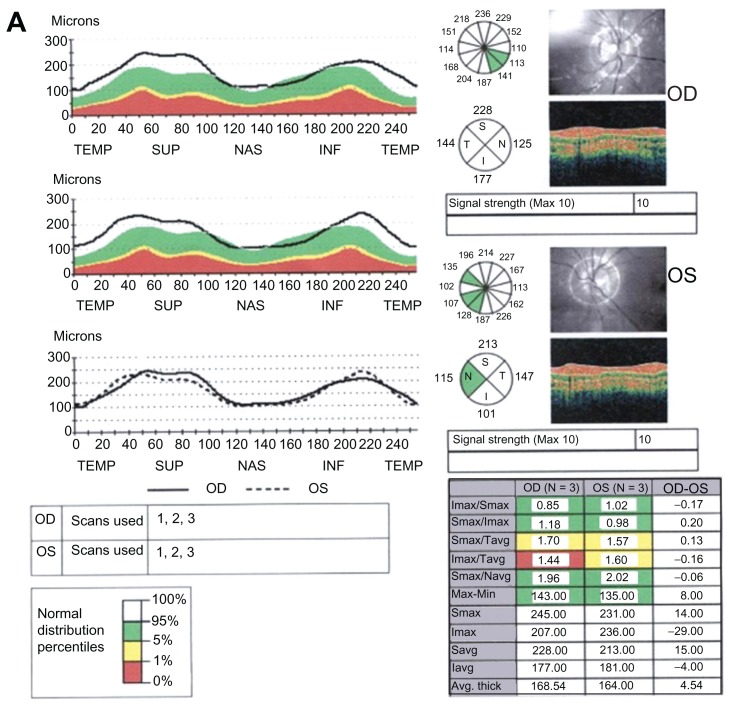
Purpose: To investigate whether or not thicker retinal nerve fiber layer (RNFL) in retinitis pigmentosa (RP) patients relates to functional abnormalities of the photoreceptors.
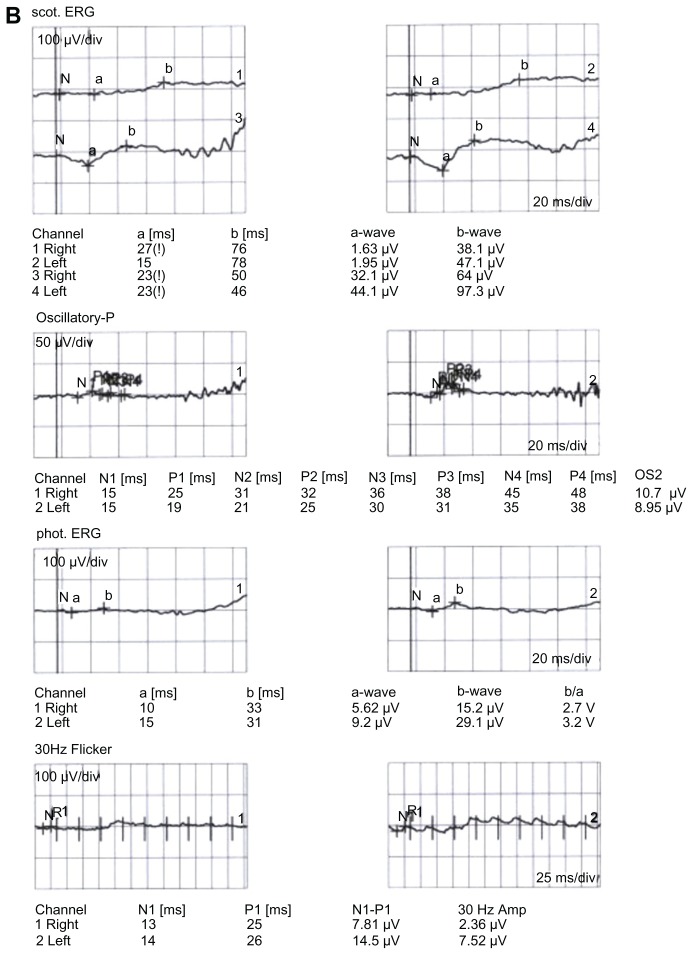
Methods: Optical coherence tomography-based RNFL thickness was measured by Stratus-3™ (Zeiss, Basel, Switzerland) optical coherence tomography and electroretinogram (ERG) recordings made using the RETI-port(®) system (Roland, Wiesbaden, Germany) in 27 patients with retinitis pigmentosa and in 30 healthy subjects.
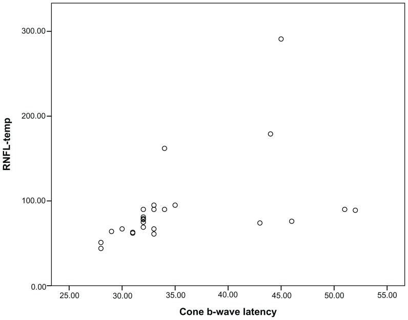
Results: Photopic ERG b-wave amplitude, cone ERG b-wave latency, 30 Hz flicker amplitude, and 30 Hz flicker latency had significant correlations to the RNFL-temporal (r = -0.55, P = 0.004, r = 0.68, P = 0.001, r = -0.65, P = 0.001, and r = -0.52, P = 0.007, respectively). Eyes with thicker RNFL (ten eyes) differed significantly from those with thinner RNFL (eight eyes) regarding cone ERG b-wave latency values only (P = 0.001).
Conclusion: Thicker RNFL in patients with retinitis pigmentosa may be associated with functional abnormality of the cone system.
Keywords: b-wave; electroretinography; optical coherence tomography; photoreceptor.
Figures
References
-
- Saloni W, Fishman GA, Edward DP, Lindeman M. Retinal nerve fiber layer defects in RP patients. Invest Ophthalmol Vis Sci. 2007;48(10):4748–4752. - PubMed
-
- Walia S, Fishman GA. Retinal nerve fiber layer analysis in RP patients using Fourier-domain OCT. Invest Ophthalmol Vis Sci. 2008;49(8):3525–3528. - PubMed
-
- Yücel Y, Gupta N. Glaucoma of the brain: a disease model for the study of transsynaptic neural degeneration. Prog Brain Res. 2008;173:465–478. - PubMed
LinkOut - more resources
Full Text Sources
