. 2012:2012:150918.
doi: 10.1155/2012/150918. Epub 2012 Mar 20.

Antiproliferative factor-induced changes in phosphorylation and palmitoylation of cytoskeleton-associated protein-4 regulate its nuclear translocation and DNA binding

Affiliations

Antiproliferative factor-induced changes in phosphorylation and palmitoylation of cytoskeleton-associated protein-4 regulate its nuclear translocation and DNA binding

David A Zacharias et al. Int J Cell Biol. 2012.

Abstract

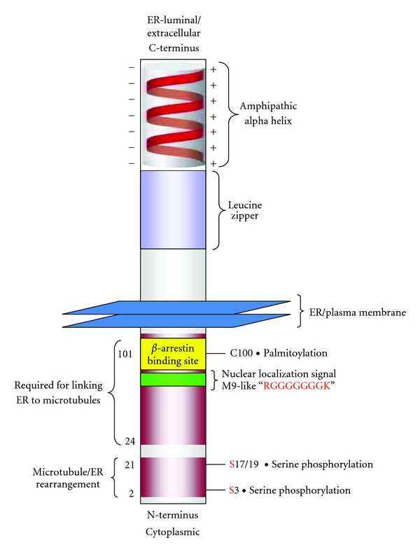
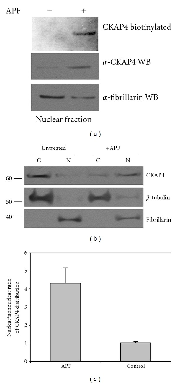
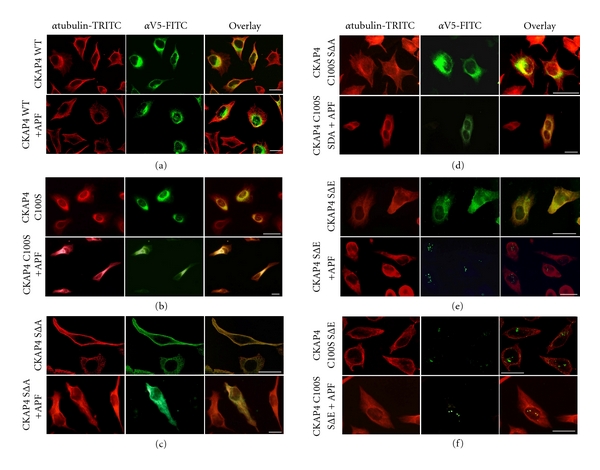
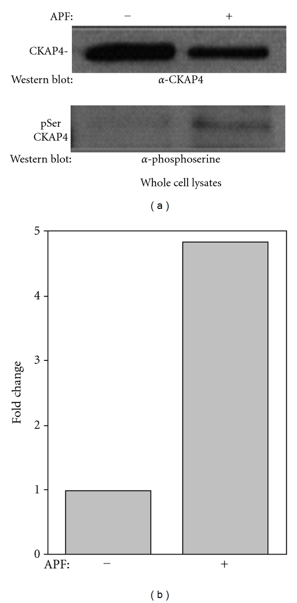
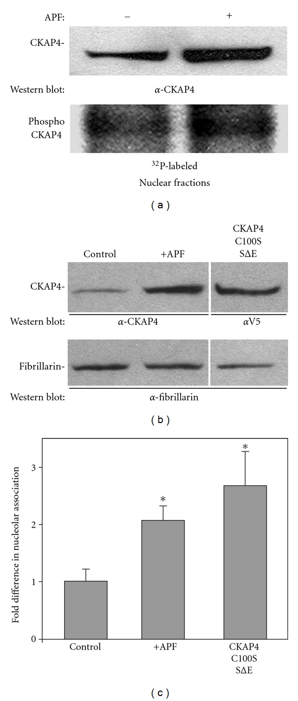
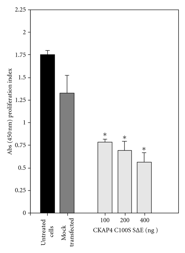
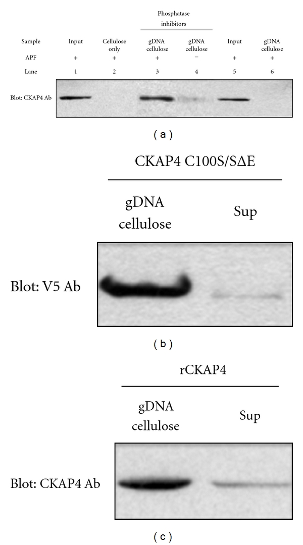
Cytoskeleton-associated protein 4 (CKAP4) is a reversibly palmitoylated and phosphorylated transmembrane protein that functions as a high-affinity receptor for antiproliferative factor (APF)-a sialoglycopeptide secreted from bladder epithelial cells of patients with interstitial cystitis (IC). Palmitoylation of CKAP4 by the palmitoyl acyltransferase, DHHC2, is required for its cell surface localization and subsequent APF signal transduction; however, the mechanism for APF signal transduction by CKAP4 is unknown. In this paper, we demonstrate that APF treatment induces serine phosphorylation of residues S3, S17, and S19 of CKAP4 and nuclear translocation of CKAP4. Additionally, we demonstrate that CKAP4 binds gDNA in a phosphorylation-dependent manner in response to APF treatment, and that a phosphomimicking, constitutively nonpalmitoylated form of CKAP4 localizes to the nucleus, binds DNA, and mimics the inhibitory effects of APF on cellular proliferation. These results reveal a novel role for CKAP4 as a downstream effecter for APF signal transduction.


Figures

Figure 1
Figure 1
CKAP4 domains. CKAP4 is a 63 kDa, oligomeric, type II, single-pass TM domain protein that is palmitoylated and phosphorylated. The luminal/extracellular domain contains an amphipathic alpha helical region (506-KVQEQVHTLLSQDQA QAARLPPQDFLDRLSSLDNLKASVSQVEADLKMLRTAVDSLVAYSV KIETNENNLESAKGLLDDLRNDLDRLFVKVEKIHEKV-602) that is important for oligomerization and a leucine zipper (468-LASTVRSLGETQLVLYGDVEELKRSVGELPSTVESL-504). Together, these regions are homologous to the DNA-binding domain of bZIP transcription factors. The cytoplasmic, N-terminus contains a cysteine residue adjacent to the TM domain at position 100 that is palmitoylated (a modification that is important for trafficking from the ER to the PM), and three serine residues (S3, S17, and S19) that are required for phosphorylation-dependent binding of CKAP4 to the microtubule cytoskeleton. Two regions in the cytoplasmic N-terminus are required for binding to and bundling microtubules, thereby maintaining the connection between the ER and the cytoskeleton [3]. There is also a PQ protein-protein interaction domain (49-PHPQQHPQQHPQNQ-63) and a putative glycine-rich nuclear localization signal sequence (65-GKGGHRGGGGGGGK-79).
Figure 2
Figure 2
Surface-labeled CKAP4 translocates from the plasma membrane into the nucleus following APF exposure. (a) HeLa cell-surface proteins were labeled with Sulfo NHS-biotin as described in Section 2. Following exposure to 20 nM APF for 24 hours (or no treatment), the cells were harvested and the nuclear protein fraction was isolated (Pierce NE-PER), separated by SDS-PAGE, and transferred to nitrocellulose. The membrane was then probed with streptavidin-HRP (1 : 5000; Pierce) to bind biotinylated proteins, and the signal was detected by ECL (Pierce). Following detection of the biotinylated proteins from the nucleus, the (streptavidin) HRP on the membrane was inactivated by incubating the blot in PBS containing 3% H2O2 and 1% sodium azide. The same membrane was then reprobed with antibodies to CKAP4 (“anti-CLIMP-63”, diluted 1 : 1000, Alexis Biochemicals) and fibrillarin (a nuclear marker and loading control; Abcam; diluted 1 : 1000). (b) HeLa cells were treated with APF (20 nM) for 24 hours, which resulted in a significant increase in the abundance of CKAP4 in the nucleus compared to control samples. Treated cells were harvested and the nuclear and cytosolic fractions were isolated and separated by SDS-PAGE as described in Section 2. Protein expression was analyzed by Western Blotting with antibodies for β-tubulin (diluted 1 : 1000, Abcam; loading control for the nonnuclear fraction), CKAP4 (“anti-CLIMP-63”, diluted 1 : 1000, Alexis Biochemicals), and fibrillarin (diluted 1 : 1000, Abcam; loading control and specific marker for the nuclear fraction), and then with an HRP-conjugated anti-mouse secondary antibody (1 : 20000; ThermoFisher Scientific). The proteins were detected by ECL (Pierce) with multiple exposures to film. The integrated density of the bands on the film was measured using ImageJ. Exposure times were controlled to ensure that the signals on film were not saturated. (c) The nuclear/cytosolic ratio represents the relative distribution of CKAP4 in the nuclear versus cytosolic fractions extracted from cells treated with or without APF. CKAP4 abundance in the APF-treated and control samples were normalized for loading to β-tubulin for the nonnuclear fractions and to fibrillarin for the nuclear fractions. The nuclear/cytosolic ratio for CKAP4 in the APF and control samples was determined from these normalized values. The standard deviation describes the variability among the normalized, nuclear, and cytosolic ratios from three independent experiments. A two tailed, paired t-test of the two data arrays (plus APF and control) indicate that the difference between these ratios is significant (P = 0.01; n = 3). Cells treated with APF stop dividing, so the 10 cm dishes containing control and APF treated cells contained fewer cells (and protein) at the end of the experiment, normalizing the CKAP4 signals to loading controls corrected for this disparity. Fibrillarin is a well-characterized nuclear marker that is also known to localize to nucleoli. The data shown are representative of four independent experiments.
Figure 3
Figure 3
CKAP4 phosphorylation and palmitoylation regulate CKAP4 trafficking. CKAP4 mutants that mimic constitutive depalmitoylation and various states of serine [3, 17, 19] phosphorylation were generated to determine the effect of these two posttranslational modifications on the subcellular distribution of CKAP4 in response to APF (see Table 1). HeLa cells were transfected for 24–36 hours with the construct indicated to the left of each panel. Cells were serum starved for 6 hours and then treated with APF (20 nM) for 18–24 hours. Subsequently, the cells were fixed in 4% buffered paraformaldehyde, permeabilized with 0.1% Triton X-100, and immunostained for (1) β-tubulin (red; TRITC) and (2) V5 (green: FITC) (as described in Section 2) to distinguish the transfected, V5-epitope tagged WT (a) and mutant versions of CKAP4 from endogenous CKAP4 (b)–(d). Mutant versions of CKAP4 that cannot be palmitoylated (C100S) or phosphorylated (SΔA) do not translocate to the nucleus in response to APF. (e) Those that mimic phosphorylation (SΔE) translocate to the nucleus in response to APF. (f) CKAP4C100SSΔE, which is constitutively depalmitoylated and phosphomimicking, is expressed primarily in the nucleus. Images taken in each channel were superimposed to illustrate the distribution of mutant CKAP4 with respect to the cytoskeleton. The cells were imaged by epifluorescence or confocal microscopy at 60X and 63X, respectively, (Scale bars = 25 microns).
Figure 4
Figure 4
APF induces serine phosphorylation of CKAP4. (a) APF treatment induces a significant increase in serine phosphorylation of CKAP4 as demonstrated by immunoprecipitation of CKAP4 followed immunoblotting to detect phosphoserine. Whole cell lysates (500 μg) from HeLa cells treated with 20 nM APF or serum-starved controls were immunoprecipitated with CKAP4 antibody (Alexis) overnight at 4°C. Samples were then bound to Protein A, washed (4X with RIPA/Empigen buffer), eluted (4X LDS sample buffer; Invitrogen), boiled at 95°C for 5 min and resolved on a 4–12% Bis-Tris gel and transferred to a nitrocellulose membrane under reducing conditions. Western Blot analysis for pSer detected phospho-serine using primary (Invitrogen and secondary antibodies (goat anti-rabbit HRP-labeled antibody; Pierce)) developed with Enhanced Chemiluminescence reagent (Pierce) and exposed to film. The membrane was stripped with Restore Stripping Buffer (Pierce) and reprobed for CKAP4 (Alexis) to normalize the phosphoserine signal to the amount of immunoprecipitated CKAP4. (b) Densitometric analysis of the immunoreactive bands was done using ImageJ, and the ratios of phosphorylated to nonphosphorylated CKAP4 were determined.
Figure 5
Figure 5
Nuclear CKAP4 is phosphorylated following APF-induced translocation. (a) CKAP4 in the nuclear fraction was phosphorylated to the same degree in APF-treated and control cells as demonstrated by metabolic labeling with γ 32P-ATP. The ratio of the CKAP4 phosphorylation signal over the corresponding CKAP4 Western Blot signal was approximately the same for both (1.00 versus 0.975; APF treated versus control). HeLa cells at ~80% confluence in 10 cm dishes were serum starved for 3 hours then 150 μCi γ 32P-ATP was added to each dish for 1 additional hour. Then the cells were either exposed to APF (20 nM; 24 hours) or left in serum-free medium (control) for 24 hours. At the end of 24 hours, the cells were harvested, washed three times in ice-cold PBS, and the nuclear and cytoplasmic fractions were isolated using the NE-PER (Pierce). Equal quantities of each fraction were separated by SDS-PAGE and transferred to nitrocellulose. The membrane was incubated overnight at 4°C in α-CKAP4 antibody (1 : 500) in TBST and 1% milk, washed and incubated for six hours at 4°C in a-mouse, HRP-conjugated secondary antibody (Pierce; 1 : 20,000) in TBST and 1% milk. CKAP4 bands were detected by enhanced chemiluminescence (ECL; Pierce; 20 second exposure; left panel). Following the Western Blot, the membrane was rinsed in 1% H2O2 for 1 minute to eliminate the chemiluminescent signal then wrapped in plastic wrap and exposed to film for 12 hours to detect the phosphorylated proteins. Densitometric analysis of the immunoreactive bands was done using ImageJ, and the ratios of phosphorylated to nonphosphorylated CKAP4 were determined. APF increases the association of CKAP4 with nucleoli. (b) and (c) Western Blot analysis shows that treatment of HeLa cells with APF (20 nM) increased the association of endogenous CKAP4 with the nucleolar fraction, and that the constitutively depalmitoylated and phosphomimicking CKAP4 mutant, CKAP4C100S/SΔE, associated with the nucleolar fraction to a greater extent than endogenous CKAP4 isolated from APF-treated cells. Nucleoli were isolated using a variation on the method published by Busch and coworkers [20] as described by the Angus Lamond lab (University of Dundee, UK). The nucleolar proteins were separated by SDS-PAGE and Western Blotted for CKAP4 (α-V5 in the case of CKAP4C100S/SΔE) and fibrillarin. The bands were detected on film by ECL and quantified using ImageJ. The CKAP4 signal in each lane of the Western Blot was normalized to the fibrillarin band in the same lane. The normalized value for CKAP4 from control cells was set to 1 and the other values were set relative to control. The values in the graph are means and SD. “*” indicates that the means of the values were significantly different than serum-starved control when evaluated using the students t-test (two-tailed. Serum starved versus APF treated P = 0.018; serum starved versus CKAP4C100S/SΔE P = 0.002; CKAP4C100S/SΔE versus APF treated, P = 0.008; n = 3 for each).
Figure 6
Figure 6
CKAP4C100S/SΔE expression inhibits HeLa cell proliferation. HeLa cells were mock transfected (Fugene; control) or transfected with 100, 200, or 400 ng of CKAP4C100S/SΔE DNA. After an additional 24 hours of growth, proliferation was measured using the WST-1 proliferation assay (Biovision) according to the manufacturer's protocol. Each data point is the mean and SD from 12 independent wells. Statistically significant differences in the rate of cellular proliferation were detected for HeLa cells transfected with 100, 200, and 400 ng of CKAP4C100S/SΔE DNA versus mock-transfected cells using the students t-test (*P < 0.001).
Figure 7
Figure 7
CKAP4 binds directly to genomic DNA. (a) CKAP4 binds gDNA in an APF-dependent manner (lane 3) when compared to mock-treated control (lane 4). The CKAP4-gDNA binding is phosphorylation dependent as CKAP4 isolated from cells treated without phosphatase inhibitors prior to APF treatment failed to bind gDNA (lane 6). As controls, we loaded APF treated nuclear lysates in lane 1 and phosphatase/APF treated lysates in lane 5. Lane 2 is a no gDNA cellulose control. (b) The ability of transiently transfected V5-tagged, CKAP4 C100S/SΔE to bind gDNA in the absence of APF treatment (c) and the ability of purified rCKAP4 (residues 126–501, which includes the bZIP-like DNA-binding domain) to bind gDNA were also assessed by comparing the amount of CKAP4 captured relative to what remained in the cell lysate after binding (Sup).

References

    1. Mundy DI, Warren G. Mitosis and inhibition of intracellular transport stimulate palmitoylation of a 62-kD protein. Journal of Cell Biology. 1992;116(1):135–146. - PMC - PubMed
    1. Klopfenstein DRC, Kappeler F, Hauri HP. A novel direct interaction of endoplasmic reticulum with microtubules. EMBO Journal. 1998;17(21):6168–6177. - PMC - PubMed
    1. Schweizer A, Rohrer J, Hauri HP, Kornfeld S. Retention of p63 in an ER-Golgi intermediate compartment depends on the presence of all three of its domains and on its ability to form oligomers. Journal of Cell Biology. 1994;126(1):25–39. - PMC - PubMed
    1. Schweizer A, Rohrer J, Jeno P, De Maio A, Buchman TG, Hauri HP. A reversibly palmitoylated resident protein (p63) of an ER-Golgi intermediate compartment is related to a circulatory shock resuscitation protein. Journal of Cell Science. 1993;104(3):685–694. - PubMed
    1. Schweizer A, Ericsson M, Bachi T, Griffiths G, Hauri HP. Characterization of a novel 63 kDa membrane protein. Implications for the organization of the ER-to-Golgi pathway. Journal of Cell Science. 1993;104(3):671–683. - PubMed