. 2012:2012:512721.
doi: 10.1155/2012/512721. Epub 2012 Mar 27.

Pexophagy: the selective degradation of peroxisomes

Affiliations

Pexophagy: the selective degradation of peroxisomes

Andreas Till et al. Int J Cell Biol. 2012.

Abstract

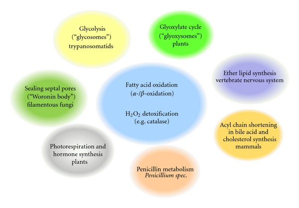
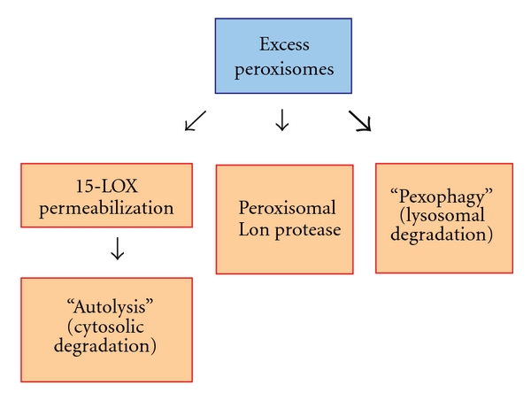
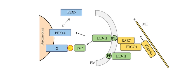
Peroxisomes are single-membrane-bounded organelles present in the majority of eukaryotic cells. Despite the existence of great diversity among different species, cell types, and under different environmental conditions, peroxisomes contain enzymes involved in β-oxidation of fatty acids and the generation, as well as detoxification, of hydrogen peroxide. The exigency of all eukaryotic cells to quickly adapt to different environmental factors requires the ability to precisely and efficiently control peroxisome number and functionality. Peroxisome homeostasis is achieved by the counterbalance between organelle biogenesis and degradation. The selective degradation of superfluous or damaged peroxisomes is facilitated by several tightly regulated pathways. The most prominent peroxisome degradation system uses components of the general autophagy core machinery and is therefore referred to as "pexophagy." In this paper we focus on recent developments in pexophagy and provide an overview of current knowledge and future challenges in the field. We compare different modes of pexophagy and mention shared and distinct features of pexophagy in yeast model systems, mammalian cells, and other organisms.


Figures

Figure 1
Figure 1
Overview of peroxisome functions in different organisms and tissues. Peroxisomes display a great variety in metabolic pathways as defined by their respective enzymatic content. Most eukaryotes share peroxisomal enzymes for fatty-acyl-CoA metabolism (α- and β-oxidation) and detoxification of hydrogen peroxide by catalase. In addition, several specialized metabolic pathways housed in the peroxisomal matrix of various organisms or tissues are shown.
Figure 2
Figure 2
Comparison of peroxisome number and morphology in different eukaryotic cells and under different proliferation conditions. (a) Upper panel: Human HeLa cells expressing the peroxisomal marker, RFP-SKL, under basal growth conditions. Lower panel: S. cerevisiae cells expressing RFP-SKL after peroxisome induction in oleate medium. The relative number of peroxisomes per cell differs greatly between different eukaryotic cell types. Size marker = 2 μm. (b) Grayscale images of P. pastoris cells expressing BFP-SKL as peroxisomal marker. Upper panel: large, clustered methanol-induced peroxisomes; lower panel: small, unclustered oleate-induced peroxisomes. Note the difference in size and appearance of peroxisomes induced by different carbon sources. Size marker = 2 μm.
Figure 3
Figure 3
Similarities and differences between selective autophagy pathways. Various selective autophagy pathways share similar molecular mechanisms. They require a receptor that interacts with the cargo, recruits a scaffold protein (Atg11) that organizes the core autophagic machinery at the PAS, and mediates recruitment of Atg8, which initiates phagophore elongation from the PAS. In the Cvt pathway (a) Atg19 and Atg34 are the receptors for the cargo proteins aminopeptidase I (Ape1) and alpha-mannosidase, respectively. These receptors bind to Atg11 at the Cvt-specific PAS to initiate membrane expansion of the phagophore. (b) The mitophagy-specific phagophore membrane expansion from the PAS is initiated by Atg32, a mitochondrial outer membrane protein. Atg32 also interacts with Atg11 and Atg8. (c) The pexophagy receptor, Atg30, is localized at the peroxisome membrane, via interaction with the PMPs, Pex3, and Pex14. It is phosphorylated upon induction of pexophagy resulting in interaction of Atg30 with core autophagic machinery components, Atg11 and Atg17. In the case of pexophagy, the direct Atg8 interaction partner is still unknown.
Figure 4
Figure 4
Micropexophagy and macropexophagy. (a) Micropexophagy differs from macropexophagy in vacuole dynamics and formation of the MIPA instead of the pexophagosome. A pexophagy-specific PAS, required for both forms of pexophagy, is characterized by its localization near the peroxisome and also touching the vacuolar membrane. Micropexophagy can target a peroxisome cluster for degradation by vacuole remodeling to form cup-like vacuolar sequestration membranes (VSMs) and a lid-like cover called the MIPA (micropexophagy-specific membrane apparatus). Macropexophagy is characterized by individual sequestration of targeted peroxisomes into a pexophagosome, followed by its fusion with the vacuole for degradation and recycling. Pexophagy signaling is dependent on Mitogen-activated protein kinase (MAPK) pathways (Mid2-Slt2 cascade), but may also be triggered by internal (unknown) factors, including signals related to the status of, or metabolic need for, (e.g., damaged or superfluous) peroxisomes. (b) The upper panel depicts a single P. pastoris cell that has undergone peroxisome induction (in methanol) and has then been switched to micropexophagy conditions (glucose). The vacuole (red, FM 4–64) is shown surrounding the targeted peroxisome cluster (blue, BFP-SKL). The MIPA (green, GFP-Atg8) forms a lid over the cup-like VSMs. The lower panel illustrates pexophagosome formation around a single peroxisome under macropexophagy conditions (ethanol). (c) S. cerevisiae cell labeled with GFP-tagged thiolase (a peroxisome matrix marker) and vacuole marker (FM 4–64, red) shows proliferated peroxisomes under nutrient-rich conditions (in oleate, top panel). When the cells are switched to glucose without nitrogen, peroxisomes are targeted to the vacuole by macropexophagy and GFP accumulates in the vacuole (lower panel).
Figure 5
Figure 5
Signal transduction cascades regulating selective autophagy in yeast. Mitogen-activated protein kinase (MAPK) cascades contribute to differential regulation of selective autophagy pathways. As recently shown, the Slt2 and Hog1 signal transduction pathways regulate both mitophagy and pexophagy [37, 96]. Besides the obvious role of environmental factors such as nutritional conditions, details of other upstream events are poorly understood.
Figure 6
Figure 6
Peroxisome degradation pathways in mammalian cells. Surplus peroxisomes or their contents (e.g., peroxisomal matrix proteins) can be degraded by at least three distinct mechanisms: Lon protease-mediated proteolysis, 15-lipoxygenase (15-LOX)-mediated cytosolic degradation (autolysis), and pexophagy (autophagy-mediated lysosomal degradation). Current studies suggest that the majority of peroxisomes are degraded by pexophagy (indicated by bold arrow).
Figure 7
Figure 7
Hypothetical mechanistic model of pexophagy in mammalian cells. Processed and lipidated LC3 (LC3-II) is integrated into the expanding phagophore membrane (PM) and also may be involved in facilitating directed movement of the PM structure by interacting with microtubules (MT) via the RAB7 effector FYCO1 and motor protein Kinesin. Targeting of peroxisomes may either be accomplished by p62-mediated detection of ubiquitin (UB) motifs on still unknown peroxisomal membrane (or membrane associated) proteins (X) or by direct binding of LC3 to PEX14, a process which is discussed to compete with the binding of PEX5 to PEX14 (dotted arrow). See text for details.

References

    1. Rhodin J. Correlation of ultrastructural organization and function in normal and experimentally changed proximal tubule cells of the mouse kidney. Stockholm, Sweden: Karolinska Institute; 1954. Ph.D. thesis.
    1. De Duve C, Baudhuin P. Peroxisomes (microbodies and related particles) Physiological Reviews. 1966;46(2):323–357. - PubMed
    1. Hayashi M, Toriyama K, Kondo M, et al. Functional transformation of plant peroxisomes. Cell Biochemistry and Biophysics. 2000;32:295–304. - PubMed
    1. Michels PAM, Moyersoen J, Krazy H, Galland N, Herman M, Hannaert V. Peroxisomes, glyoxysomes and glycosomes. Molecular Membrane Biology. 2005;22(1-2):133–145. - PubMed
    1. Moyersoen J, Choe J, Fan E, Hol WGJ, Michels PAM. Biogenesis of peroxisomes and glycosomes: trypanosomatid glycosome assembly is a promising new drug target. FEMS Microbiology Reviews. 2004;28(5):603–643. - PubMed

LinkOut - more resources