

The OSCAR-IB consensus criteria for retinal OCT quality assessment
- PMID: 22536333
- PMCID: PMC3334941
- DOI: 10.1371/journal.pone.0034823
The OSCAR-IB consensus criteria for retinal OCT quality assessment
Abstract
Background: Retinal optical coherence tomography (OCT) is an imaging biomarker for neurodegeneration in multiple sclerosis (MS). In order to become validated as an outcome measure in multicenter studies, reliable quality control (QC) criteria with high inter-rater agreement are required.
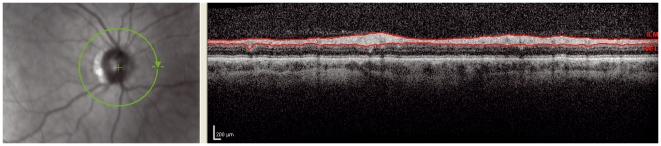
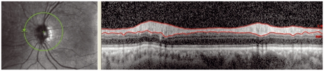
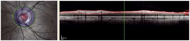
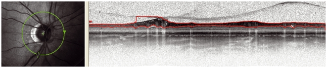
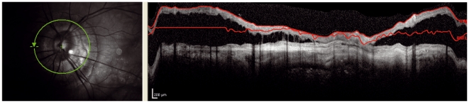
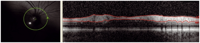
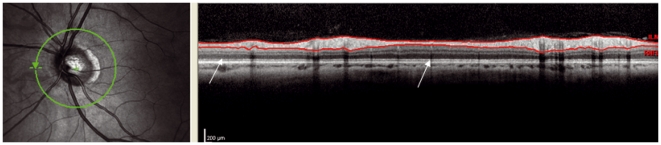
Methods/principal findings: A prospective multicentre study on developing consensus QC criteria for retinal OCT in MS: (1) a literature review on OCT QC criteria; (2) application of these QC criteria to a training set of 101 retinal OCT scans from patients with MS; (3) kappa statistics for inter-rater agreement; (4) identification reasons for inter-rater disagreement; (5) development of new consensus QC criteria; (6) testing of the new QC criteria on the training set and (7) prospective validation on a new set of 159 OCT scans from patients with MS. The inter-rater agreement for acceptable scans among OCT readers (n = 3) was moderate (kappa 0·45) based on the non-validated QC criteria which were entirely based on the ophthalmological literature. A new set of QC criteria was developed based on recognition of: (O) obvious problems, (S) poor signal strength, (C) centration of scan, (A) algorithm failure, (R) retinal pathology other than MS related, (I) illumination and (B) beam placement. Adhering to these OSCAR-IB QC criteria increased the inter-rater agreement to kappa from moderate to substantial (0.61 training set and 0.61 prospective validation).
Conclusions: This study presents the first validated consensus QC criteria for retinal OCT reading in MS. The high inter-rater agreement suggests the OSCAR-IB QC criteria to be considered in the context of multicentre studies and trials in MS.
Conflict of interest statement
Figures
References
-
- Frohman E, Costello F, Zivadinov R, Stuve O, Conger A, et al. Optical coherence tomography in multiple sclerosis. Lancet Neurol. 2006;5:853–863. - PubMed
-
- Petzold A, de Boer J, Schippling S, Vermersch P, Kardon R, et al. Optical coherence tomography in multiple sclerosis: a systematic review and meta-analysis. Lancet Neurol. 2010;9:921–932. - PubMed
-
- Jindahra P, Hedges TH, Mendoza-Santiesteban CE, Plant GT. Optical coherence tomography of the retina: applications in neurology. Curr Opin Neurol. 2010;23:16–23. - PubMed
-
- Barkhof F, Calabresi PA, Miller DH, Reingold SC. Imaging outcomes for neuroprotection and repair in multiple sclerosis trials. Nat Rev Neurol. 2009;5:256–266. - PubMed
Publication types
MeSH terms
LinkOut - more resources
Full Text Sources
Medical
