. 2012;7(4):e35783.
doi: 10.1371/journal.pone.0035783. Epub 2012 Apr 20.

Tau phosphorylation and μ-calpain activation mediate the dexamethasone-induced inhibition on the insulin-stimulated Akt phosphorylation

Affiliations

Tau phosphorylation and μ-calpain activation mediate the dexamethasone-induced inhibition on the insulin-stimulated Akt phosphorylation

Yudong Liu et al. PLoS One. 2012.

Abstract

Evidence has suggested that insulin resistance (IR) or high levels of glucocorticoids (GCs) may be linked with the pathogenesis and/or progression of Alzheimer's disease (AD). Although studies have shown that a high level of GCs results in IR, little is known about the molecular details that link GCs and IR in the context of AD. Abnormal phosphorylation of tau and activation of μ-calpain are two key events in the pathology of AD. Importantly, these two events are also related with GCs and IR. We therefore speculate that tau phosphorylation and μ-calpain activation may mediate the GCs-induced IR. Akt phosphorylation at Ser-473 (pAkt) is commonly used as a marker for assessing IR. We employed two cell lines, wild-type HEK293 cells and HEK293 cells stably expressing the longest human tau isoform (tau-441; HEK293/tau441 cells). We examined whether DEX, a synthetic GCs, induces tau phosphorylation and μ-calpain activation. If so, we examined whether the DEX-induced tau phosphorylation and μ-calpain activation mediate the DEX-induced inhibition on the insulin-stimulated Akt phosphorylation. The results showed that DEX increased tau phosphorylation and induced tau-mediated μ-calpain activation. Furthermore, pre-treatment with LiCl prevented the effects of DEX on tau phosphorylation and μ-calpain activation. Finally, both LiCl pre-treatment and calpain inhibition prevented the DEX-induced inhibition on the insulin-stimulated Akt phosphorylation. In conclusion, our study suggests that the tau phosphorylation and μ-calpain activation mediate the DEX-induced inhibition on the insulin-stimulated Akt phosphorylation.


Conflict of interest statement

Competing Interests: The authors have declared that no competing interests exist.

Figures

Figure 1
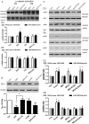
Figure 1. Effects of dexamethasone on the insulin-stimulated Akt phosphorylation.
A): Both wild-type HEK293 and HEK293/tau441 cells were treated with 1 µM dexamethasone (DEX) for 1–6 days and then stimulated with 100 nM insulin (i) for 15 min. Representative immunoblots from the results on the third day (D3) and the sixth day (D6) after DEX treatment were shown. Bars representing means ± SEM were shown below. Total amounts of Akt remained stable under the conditions. In HEK293/tau441 cells, DEX prevented the insulin-stimulated increases in pAkt on D3 and D6. In wild-type HEK293 cells, the inhibitory effect of DEX was evident on D6 but not on D3. Each experiment was repeated three times unless stated otherwise. * P<0.05 versus control. B): Culture cells were pre-treated with mifepristone (RU; 20 µM, 30 min) and then treated with DEX for 3 days or 6 days. Representative immunoblots from wild-type HEK293 cells on D6 and those from HEK293/tau441 cells on D3 were shown, with bars representing means ± SEM below. Pre-treatment with RU prevented the inhibitory effects of DEX. * P<0.05 versus DEX group.
Figure 2
Figure 2. Effects of DEX and lithium chloride on tau phosphorylation.
HEK293/tau441 cells were pre-treated with lithium chloride (LiCl; 10 mM, 1 h) and then treated with DEX for 3 days (D3) or 6 days (D6), followed by Western blotting analysis of tau phosphorylation with Tau-1 (against dephosphorylated tau within the epitope 189–207) and R134d (against total tau) antibodies. Total amounts of tau remained largely stable and the level of tau phosphorylation was determined by the Tau-1/R134d ratio. Bars representing means ± SEM. On D3, DEX induced an increase in tau phosphorylation, as evidenced by the decrease in the Tau-1/R134d ratio. Pre-treatment with LiCl prevented the effect of DEX on tau phosphorylation on D3. * P<0.05 versus control, # P<0.05 between indicated groups.
Figure 3
Figure 3. Effects of insulin on tau phosphorylation.
HEK293/tau441 cells were pre-treated with LiCl (10 mM, 1 h) and then treated with DEX for 3 days, followed by stimulation of insulin for 15 min and Western analysis of Tau-1 and R134d. Bars representing means ± SEM. Insulin did not prevent the DEX-induced decrease in Tau-1 or affected Tau-1 levels under other conditions, suggesting that insulin does not affect tau phosphorylation. * P<0.05 versus control.
Figure 4
Figure 4. Effects of LiCl on the DEX-induced inhibitory effect.
Both wild-type HEK293 and HEK293/tau441 cells were pre-treated with LiCl (10 mM, 1 h) and then treated with DEX for 3 days (D3; A) or 6 days (D6; B), followed by stimulation of insulin and Western analysis of pAkt and Akt. Bars representing means ± SEM. Pre-treatment with LiCl prevented the inhibitory effect of DEX in HEK293/tau441 cells on D3.
Figure 5
Figure 5. Involvement of μ-calpain in the inhibitory effect of DEX.
A): Both wild-type HEK293 and HEK293/tau441 cells were pre-treated with LiCl (10 mM, 1 h) and then treated with DEX for 3 days (D3) or 6 days (D6). Activation of μ-calpain was determined by the ratios of the active/truncated calpain (78-kDa bands) and the inactive/full-length calpain (80-kDa bands). Bars representing means ± SEM. On D3 in HEK293/tau441 cells, DEX induced μ-calpain activation and pre-treatment with LiCl prevented the activation of μ-calpain. * P<0.05 versus control. B): HEK293/tau441 cells were pre-treated with E-64d (30 µg/ml, 1 h) or LiCl (10 mM, 1 h), and then treated with DEX for 3 days. The level of tau phosphorylation was determined by the ratio of Tau-1/R134d. Bars representing means ± SEM. E-64d did not have obvious effect on the DEX-induced increase in tau phosphorylation. * P<0.05 versus control, # P<0.05 between indicated groups. C) Both wild-type HEK293 and HEK293/tau441 cells were pre-treated with E-64d (30 µg/ml, 1 h) and then treated with DEX for 3 days (D3) or 6 days (D6), followed by stimulation of insulin and Western analysis of pAkt and Akt. Bars representing means ± SEM. Pre-treatment with E-64d prevented the inhibitory effect of DEX in HEK293/tau441 cells on D3. * P<0.05 versus control.

References

    1. Reaven GM. Insulin resistance: the link between obesity and cardiovascular disease. Med Clin North Am. 2011;95:875–892. - PubMed
    1. Tsoyi K, Jang HJ, Nizamutdinova IT, Park K, Kim YM, et al. PTEN differentially regulates expression of ICAM-1 and VCAM-1 through PI3K/Akt/GSK-3β/GATA-6 signaling pathways in TNF-α-activated human endothelial cells. Atherosclerosis. 2010;213:115–121. - PubMed
    1. Lu FP, Lin KP, Kuo HK. Diabetes and the risk of multi-system aging phenotypes: a systematic review and meta-analysis. PLoS One. 2009;4:e4144. - PMC - PubMed
    1. Park SA. A common pathogenic mechanism linking type-2 diabetes and Alzheimer's disease: evidence from animal models. J Clin Neurol. 2011;7:10–18. - PMC - PubMed
    1. Baker LD, Cross DJ, Minoshima S, Belongia D, Watson GS, et al. Insulin resistance and Alzheimer-like reductions in regional cerebral glucose metabolism for cognitively normal adults with prediabetes or early type 2 diabetes. Arch Neurol. 2011;68:51–57. - PMC - PubMed

Publication types

MeSH terms