. 2012 Jun 15;366(2):298-307.
doi: 10.1016/j.ydbio.2012.03.015. Epub 2012 Apr 17.

The Nkx5/HMX homeodomain protein MLS-2 is required for proper tube cell shape in the C. elegans excretory system

Affiliations

The Nkx5/HMX homeodomain protein MLS-2 is required for proper tube cell shape in the C. elegans excretory system

Ishmail Abdus-Saboor et al. Dev Biol. .

Abstract

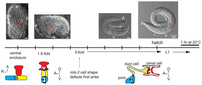
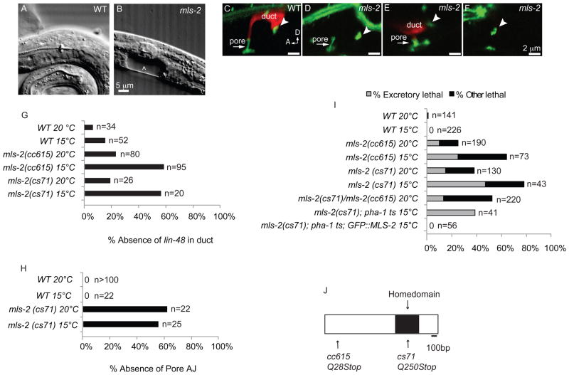
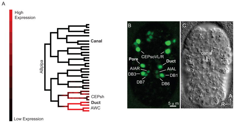
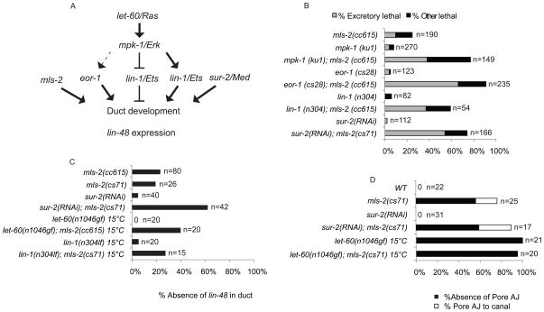
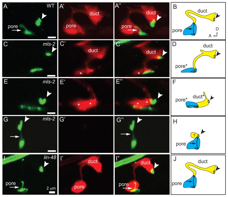
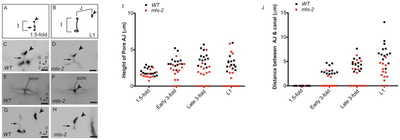
Cells perform wide varieties of functions that are facilitated, in part, by adopting unique shapes. Many of the genes and pathways that promote cell fate specification have been elucidated. However, relatively few transcription factors have been identified that promote shape acquisition after fate specification. Here we show that the Nkx5/HMX homeodomain protein MLS-2 is required for cellular elongation and shape maintenance of two tubular epithelial cells in the C. elegans excretory system, the duct and pore cells. The Nkx5/HMX family is highly conserved from sea urchins to humans, with known roles in neuronal and glial development. MLS-2 is expressed in the duct and pore, and defects in mls-2 mutants first arise when the duct and pore normally adopt unique shapes. MLS-2 cooperates with the EGF-Ras-ERK pathway to turn on the LIN-48/Ovo transcription factor in the duct cell during morphogenesis. These results reveal a novel interaction between the Nkx5/HMX family and the EGF-Ras pathway and implicate a transcription factor, MLS-2, as a regulator of cell shape.


Figures

Figure 1
Figure 1. Timeline of excretory system development
Schematics of excretory canal cell (red), duct (yellow), and pore (blue) at different developmental stages. DIC images correspond to developmental stage listed on timeline; colored circles on the DIC images represent positions of the canal, duct, and pore. Dark black lines indicate apical junctions. Dotted line, duct auto-fusion. Arrow, pore autocellular junction. Arrowhead, duct-canal cell intercellular junction. Schematics are modified from (Abdus-Saboor et al., 2011). EGF-Ras-ERK-dependent duct vs. pore cell fate specification occurs just prior to the 1.5-fold stage (Abdus-Saboor et al., 2011). The duct elongates extensively between the 1.5-fold and early 3-fold stages (Stone et al., 2009).
Figure 2
Figure 2. mls-2 mutants have incompletely penetrant and cold sensitive lethal excretory system defects
(A) WT L1 stage larva. (B) mls-2(cs71) L1 stage larva showing fluid accumulation near duct and pore (arrowhead). (C-F) L1 worms with AJM-1::GFP junction marker and lin-48p::mcherry duct marker. Arrowhead, duct-canal cell intercellular junction. (C) WT larva with one duct and one pore. (D) mls-2 mutant with normal AJM-1::GFP but no lin-48 expression. (E) mls-2 mutant with normal lin-48 duct expression and collapsing pore. (F) mls-2 mutant with no lin-48 duct expression and no pore autocellular junction. (G,H) Quantification of marker loss phenotypes in mls-2 mutants. AJ, autocellular junction. (I) Rod-like (excretory) lethality shown as a fraction of total lethality. Other lethality scored as any worm that failed to reach L4 within 4 days. Note: GFP::MLS-2 rescue data scored by DIC microscopy looking for presence or absence of fluid cysts in L1s. Non-transgenic siblings were scored as controls. Complete genotype in this experiment was: mls-2(cs71); pha-1(e2123); Ex[GFP::MLS-2; pha-1(+)]. (J) Protein structure of MLS-2 showing the homeodomain and mutant alleles. cs71 changes a CAA codon (Q250) to TAA (stop).
Figure 3
Figure 3. mls-2 is expressed in the duct and pore cell lineages
(A) Lineage tree showing florescence intensity of GFP::MLS-2 expression from 3D automated lineage analysis. Only 1 of 3 embryos that were lineaged is shown here. Only the ABplpa lineage is shown, but GFP::MLS-2 is symmetrically expressed in the ABprpa lineage, which gives rise to the excretory pore. See Supplemental Fig.1 for complete lineage analysis of all 3 embryos. (B) Ventral enclosure embryo expressing GFP::MLS-2, and (C) corresponding DIC image. Identities of some nuclei are indicated. CEPsh nuclei are dim and not visible at this stage. AWC nuclei are not in plane of focus. The pair of nuclei directly above the CEPsoVL/R nuclei are the sisters of the CEPsoVL/R that are fated to die (Sulston et al., 1983). The DB1/DB3 ventral cord motor neurons are sisters of the duct and pore, respectively. AIAL/R are amphid inter-neurons and DB6/DB7 are ventral cord motor neurons; expression in these cells initiates at this stage and is very faint.
Figure 4
Figure 4. mls-2 cooperates with the EGF-Ras-ERK pathway to promote duct differentiation and lin-48/ovo expression
(A) EGF-Ras-ERK signaling pathway downstream of let-60/Ras. eor-1, lin-1/ETS, and sur-2/Med23 are downstream nuclear effectors. mls-2 acts downstream or parallel to the EGF-Ras-ERK pathway. (B) Rod-like (excretory) lethality shown as a fraction of total lethality. Other lethality scored as any worm that failed to reach L4 within 4 days. Experiments performed at 20°C. (C) Percentage of L1 worms that lacked lin-48p::GFP expression in duct. Note: let-60(n1046) and lin-1(n304) worms frequently had two lin-48p::GFP positive nuclei instead of one. (D) Percentage of L1 worms that lacked a pore autocellular junction and worms that had a pore AJ junction connected directly to the canal cell (Pore AJ to canal), as scored with AJM-1::GFP. Note: let-60(n1046) and let-60(n1046); mls-2(cs71) were scored at late 3-fold instead of L1. mls-2 alone and in combination with sur-2 (RNAi) assessed at 20°C. let-60(n0146) alone and in combination with mls-2 assessed at 15°C. Note: eor-1(cs28) (Rocheleau et al., 2002) and lin-1(n304)(Beitel et al., 1995) are null alleles; mpk-1(ku1)(Lackner and Kim, 1998) is a hypomorphic allele. Experiments performed at 20°C unless otherwise indicated.
Figure 5
Figure 5. mls-2 affects duct and pore tube shape
AJM-1::GFP (left column) and dct-5p::mCherry (second column) in early L1s grown at 15°C, lateral view. Third column shows overlay. Fourth column shows schematic interpretation of phenotypes. Arrowhead, duct-canal cell intercellular junction. (A,B) WT; n=20. (C–H) mls-2(cs71); n=25. (C,D) Pore cell collapsed and duct extending ventrally; n=11/25. Note: In both wildtype and mls-2 mutants, the duct sometimes displays a dorsal extension as seen here. (E,F) Both the duct and pore cells collapsed ventrally; n=3/25 (G,H) Pore cell AJ connected to canal cell, with duct cell presumably small or mispositioned; n=5/25. The remaining 6/25 mls-2 mutants looked similar to WT. Asterisks indicate ventral cells with collapsed cell shapes. (I, J) lin-48(sa469); n=15. Duct cell shape appears similar to wild-type. Note: lin-48(sa469) is a strong loss-of-function allele that alters a histidine in the second C2H2 zinc finger (Chamberlin et al., 1999).
Figure 6
Figure 6. mls-2 cell shape defects begin around the elongation stage of embryogenesis
(A, B) Schematics of wild-type AJM-1::GFP junction pattern at 1.5 fold (A) and late 3-fold (B) stages. Parameters measured in I, J are indicated with brackets. Arrowhead, duct-canal cell intercellular junction. (C–H) Excretory duct and pore junction patterns visualized with AJM-1::GFP. Images were inverted in ImageJ for clarity. (C) WT and (D) mls-2 embryos at 1.5-fold, lateral view. (E) WT and (F) mls-2 mid 3-fold embryos, ventral view. Single pore opening lies just adjacent to the G2 and W epidermal cells. Note proximity of the canal cell junction (arrowhead) to the pore opening in F. (G) WT and (H) mls-2 late 3-fold embryos, lateral view. Note no duct elongation in H. (I, J) Measurements of AJ height and distance between AJ and canal cell junction at different time-points. Note: at 1.5-fold stage, height of AJ is duct and pore, and at all other stages, height of AJ is only the pore. Each point is a measurement from a single worm. Early 3-fold corresponds to 2 hours after the 1.5-fold stage. Late 3-fold corresponds to 6 hours after the 1.5-fold stage.

Similar articles

Cited by

References

    1. Abdus-Saboor I, Mancuso VP, Murray JI, Palozola K, Norris C, Hall DH, Howell K, Huang K, Sundaram MV. Notch and Ras promote sequential steps of excretory tube development in C. elegans. Development. 2011;138:3545–55. - PMC - PubMed
    1. Andrew DJ, Ewald AJ. Morphogenesis of epithelial tubes: Insights into tube formation, elongation, and elaboration. Dev Biol. 2010;341:34–55. - PMC - PubMed
    1. Bao Z, Murray JI, Boyle T, Ooi SL, Sandel MJ, Waterston RH. Automated cell lineage tracing in Caenorhabditis elegans. Proc Natl Acad Sci U S A. 2006;103:2707–12. - PMC - PubMed
    1. Bargmann CI, Hartwieg E, Horvitz HR. Odorant-selective genes and neurons mediate olfaction in C. elegans. Cell. 1993;74:515–27. - PubMed
    1. Beitel GJ, Tuck S, Greenwald I, Horvitz HR. The Caenorhabditis elegans gene lin-1 encodes an ETS-domain protein and defines a branch of the vulval induction pathway. Genes Dev. 1995;9:3149–62. - PubMed

Publication types

MeSH terms