. 2012 Jul;33(21):5451-8.
doi: 10.1016/j.biomaterials.2012.03.083. Epub 2012 Apr 25.

Protein polymer hydrogels by in situ, rapid and reversible self-gelation

Affiliations

Protein polymer hydrogels by in situ, rapid and reversible self-gelation

Daisuke Asai et al. Biomaterials. 2012 Jul.

Abstract

Protein-based biomaterials are an important class of materials for applications in biotechnology and medicine. The exquisite control of their composition, stereochemistry, and chain length offers unique opportunities to engineer biofunctionality, biocompatibility, and biodegradability into these materials. Here, we report the synthesis of a thermally responsive peptide polymer-based hydrogel composed of a recombinant elastin-like polypeptide (ELP) that rapidly forms a reversibly cross-linked hydrogel by the formation of intermolecular disulfide cross-links. To do so, we designed and synthesized ELPs that incorporate periodic cysteine residues (cELPs), and show that cELPs are thermally responsive protein polymers that display rapid gelation under physiologically relevant, mild oxidative conditions. Gelation of cELPs, at concentrations as low as 2.5 wt%, occurs in ≈ 2.5 min upon addition a low concentration of hydrogen peroxide (0.3 wt%). We show the utility of these hydrogels for the sustained release of a model protein in vitro, and demonstrate the ability of this injectable biomaterial to pervade tumors to maximize tumor coverage and retention time upon intratumoral injection. cELPs represent a new class of injectable reversibly cross-linked hydrogels with properties intermediate between ELP coacervates and chemically cross-linked ELP hydrogels that will find useful applications in drug delivery and tissue engineering.


Figures

Fig. 1
Fig. 1
Copper-stained SDS-PAGE gel of cELPs. All cELPs were purified successfully by inverse transition cycling (ITC) using 20 mM tris(2-carboxyethyl)phosphine. The symbols + and − indicate the presence and absence of cysteine in the ELPs, respectively.
Fig. 2
Fig. 2
Thermally responsive behavior of cELPs at 2.5 wt% in PBS. (A) Absorbance at 350 nm for all ELPs was measured under a temperature gradient of 1°C min−1 to determine the ELP inverse transition temperature (Tt). (B) Tt of uncross-linked cELPs in PBS at 2.5 wt%. The Tt was defined as the solution temperature that corresponded to the maximum in the first derivative of the absorbance (350 nm).
Fig. 3
Fig. 3
Effect of hydrogen peroxide (H2O2) on gelation. The final concentrations of cELP and H2O2 were 2.5 wt% and 0.3 wt%, respectively. (A) A mixture of cELP and H2O2 was incubated on ice for 3 min, and further incubated at 4°C and/or 37°C for 10 min, and the vials were then inverted to assess the fluidity of the ELP. The symbols + and − indicate the presence and absence of H2O2, respectively. (B) Thermally responsive behavior cELPs in the hydrogel state. The hydrogels were prepared in a cuvette on ice, and the resulting hydrogels were equilibrated at 15°C for 30 min before determining their turbidity profile. Data represent a typical result from three independent experiments.
Fig. 3
Fig. 3
Effect of hydrogen peroxide (H2O2) on gelation. The final concentrations of cELP and H2O2 were 2.5 wt% and 0.3 wt%, respectively. (A) A mixture of cELP and H2O2 was incubated on ice for 3 min, and further incubated at 4°C and/or 37°C for 10 min, and the vials were then inverted to assess the fluidity of the ELP. The symbols + and − indicate the presence and absence of H2O2, respectively. (B) Thermally responsive behavior cELPs in the hydrogel state. The hydrogels were prepared in a cuvette on ice, and the resulting hydrogels were equilibrated at 15°C for 30 min before determining their turbidity profile. Data represent a typical result from three independent experiments.
Fig. 4
Fig. 4
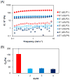
Rheological properties of cELP hydrogels. A mixture of 2.5 wt% cELP and 0.3 wt% H2O2 was loaded into the rheometer platen at 25°C, compressed to 720–740 μm, equilibrated for 5 min, and the rheological parameters were obtained by linear oscillatory frequency sweep experiments. (A) Storage modulus (G′) and loss modulus (G″) versus frequency for of cELP hydrogels. (B) Plateau modulus (G0) obtained from the plateau value of G′. Data shown are the mean ± SEM and are representative of three independent experiments.
Fig. 5
Fig. 5
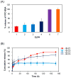
In vitro FITC-BSA release from hydrogels of cELPs. ELPs pre-mixed with FITC-BSA were mixed with H2O2 at 25°C. BSA (4 mg mL−1) was added to the resulting hydrogels, and the mixture was incubated at 37°C. The amount of released FITC-BSA was quantified by the fluorescence intensity in the supernatant. (A) Percent release of FITC-BSA after 4 h. (B) Percent cumulative release of FITC-BSA from cELP hydrogels. Each data point represents the mean value ± SEM (n = 4–7).
Fig. 6
Fig. 6
Rapid in situ-gelation of cELPs in vivo. The final concentrations of cELP and H2O2 were 2.5 wt% and 0.3 wt%, respectively for all in vivo studies. (A) Intramuscular co-injection of ELP1 with H2O2 into mice was carried out. Bromophenol blue was pre-mixed with ELP1 for visualization of the hydrogel. Three minutes after injection, the mice were incised to confirm hydrogel formation, and the hydrogel was excised from the injection site. (B) Tumor retention of ELP1 after intratumoral infusion of [125I]ELPs in a tumor-bearing mouse model. The ID%/tumor at 0, 4, 8, 24, 48, 72, 96 hour, and 1 week after administration are expressed as mean value ± SEM (n = 8–10; *, p<0.01). (C) Near-infrared in vivo imaging following intratumoral administration. The images at 8 hours after infusion are shown as pseudo-color-enhanced fluorescence intensity superimposed on the white light images. (D) Magnified images of mice 2 and 5 are shown in Fig. 6C. (E) Clearance of IRDye800CW-labelled cELP1. Fluorescence intensity in the tumor (mean pixel fluorescence) at 0, 4, 8, and 24 hour post-injection; data was expressed as mean value ± SEM (n = 4–5). *, p<0.02.
Fig. 6
Fig. 6
Rapid in situ-gelation of cELPs in vivo. The final concentrations of cELP and H2O2 were 2.5 wt% and 0.3 wt%, respectively for all in vivo studies. (A) Intramuscular co-injection of ELP1 with H2O2 into mice was carried out. Bromophenol blue was pre-mixed with ELP1 for visualization of the hydrogel. Three minutes after injection, the mice were incised to confirm hydrogel formation, and the hydrogel was excised from the injection site. (B) Tumor retention of ELP1 after intratumoral infusion of [125I]ELPs in a tumor-bearing mouse model. The ID%/tumor at 0, 4, 8, 24, 48, 72, 96 hour, and 1 week after administration are expressed as mean value ± SEM (n = 8–10; *, p<0.01). (C) Near-infrared in vivo imaging following intratumoral administration. The images at 8 hours after infusion are shown as pseudo-color-enhanced fluorescence intensity superimposed on the white light images. (D) Magnified images of mice 2 and 5 are shown in Fig. 6C. (E) Clearance of IRDye800CW-labelled cELP1. Fluorescence intensity in the tumor (mean pixel fluorescence) at 0, 4, 8, and 24 hour post-injection; data was expressed as mean value ± SEM (n = 4–5). *, p<0.02.

References

    1. Hatefi A, Amsden B. Biodegradable injectable in situ forming drug delivery systems. J Control Release. 2002;80(1–3):9–28. - PubMed
    1. Packhaeuser CB, Schnieders J, Oster CG, Kissel T. In situ forming parenteral drug delivery systems: an overview. Eur J Pharm Biopharm. 2004;58(2):445–455. - PubMed
    1. Kretlow JD, Klouda L, Mikos AG. Injectable matrices and scaffolds for drug delivery in tissue engineering. Adv Drug Deliv Rev. 2007;59(4–5):263–273. - PubMed
    1. Yu L, Ding J. Injectable hydrogels as unique biomedical materials. Chem Soc Rev. 2008;37(8):1473–1481. - PubMed
    1. Nguyen MK, Lee DS. Injectable biodegradable hydrogels. Macromol Biosci. 2010;10(6):563–579. - PubMed

Publication types

MeSH terms