. 2012 Apr 25;484(7395):479-84.
doi: 10.1038/nature11045.

Clonally dominant cardiomyocytes direct heart morphogenesis

Affiliations

Clonally dominant cardiomyocytes direct heart morphogenesis

Vikas Gupta et al. Nature. .

Abstract

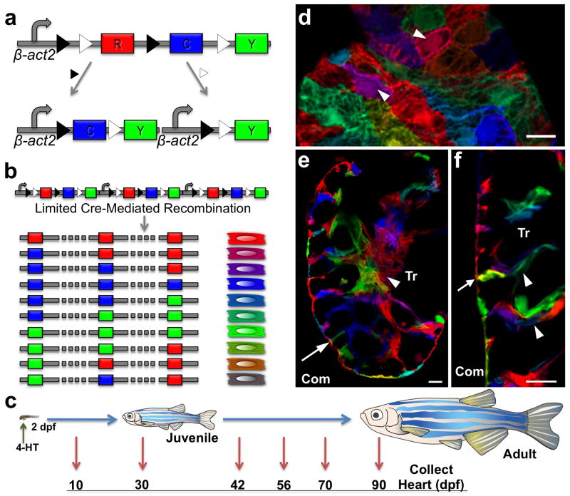
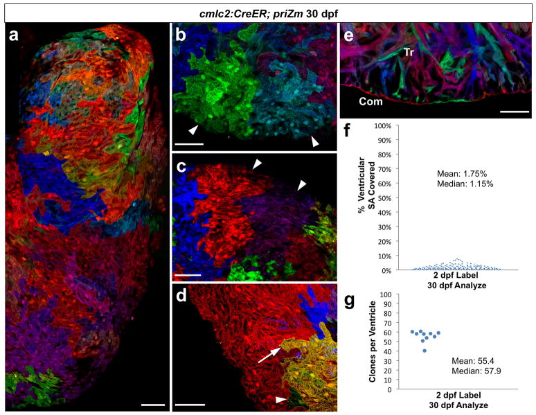
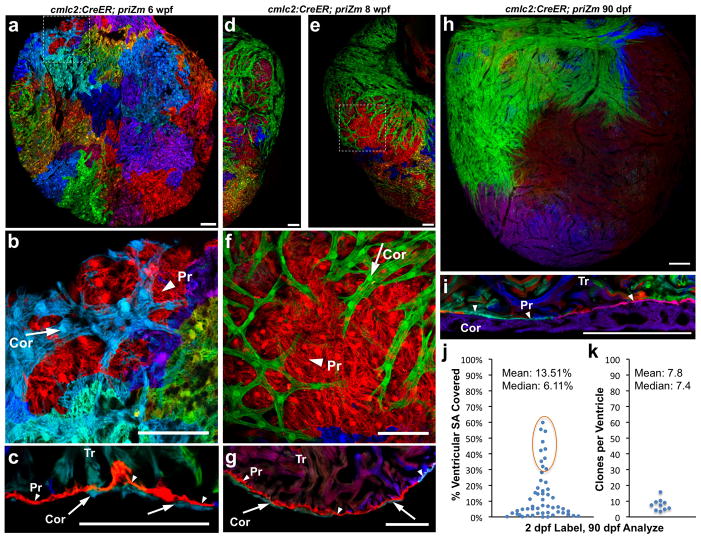
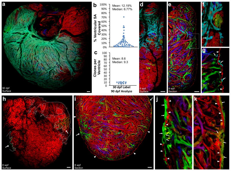
As vertebrate embryos develop to adulthood, their organs undergo marked changes in size and tissue architecture. The heart acquires muscle mass and matures structurally to fulfil increasing circulatory needs, a process that is incompletely understood. Here we used multicolour clonal analysis to define the contributions of individual cardiomyocytes as the zebrafish heart undergoes morphogenesis from a primitive embryonic structure into its complex adult form. We find that the single-cardiomyocyte-thick wall of the juvenile ventricle forms by lateral expansion of several dozen cardiomyocytes into muscle patches of variable sizes and shapes. As juvenile zebrafish mature into adults, this structure becomes fully enveloped by a new lineage of cortical muscle. Adult cortical muscle originates from a small number of cardiomyocytes--an average of approximately eight per animal--that display clonal dominance reminiscent of stem cell populations. Cortical cardiomyocytes initially emerge from internal myofibres that in rare events breach the juvenile ventricular wall, and then expand over the surface. Our results illuminate the dynamic proliferative behaviours that generate adult cardiac structure, revealing clonal dominance as a key mechanism that shapes a vertebrate organ.


Conflict of interest statement

Author Information. The authors declare no competing financial interests.

Figures

Figure 1
Figure 1. Multicolor clonal labeling of embryonic zebrafish cardiomyocytes
a, Recombination at paired lox2272 (black triangles) or loxP (white triangles) sites leads to expression of CFP or YFP, respectively. b, Limited Cre-mediated recombination of tandem cassette insertions results in combinatorial expression of fluorescent proteins. c, Cartoon of lineage-tracing experiments. d, 10 dpf ventricular surface myocardium. Single cardiomyocytes are predominantly labeled with unique colors (arrowheads). e, f, 10 dpf ventricular confocal slice, indicating trabecular cardiomyocytes connected with clonally unrelated cardiomyocytes at the wall (e, f, arrow) and within trabeculae (f, arrowheads). Com, compact muscle. Tr, trabecular muscle. Scale bars, 10 μm.
Figure 2
Figure 2. Several dozen embryonic cardiomyocytes build the juvenile ventricular wall
a, Surface myocardium of half of a 30 dpf ventricular side, displaying clonal patches of varied shapes and sizes. b, c, Cardiomyocyte clones near the apex or chamber midpoint forming wedge/stripe shapes (arrowheads). d, Single-cell clone (green, arrowhead) positioned near a large clone (yellow, arrow). e, 30 dpf ventricular confocal slice, depicting a wall of single-cardiomyocyte thickness (Com) surrounding trabecular muscle (Tr). f, %Surface area occupied by 30 dpf clones (146 clones, 5 ventricles). g, Surface clones per ventricle (n=10). Scale bars, 50 μm.
Figure 3
Figure 3. Clonally dominant cortical cardiomyocytes
a–c, 6 weeks post-fertilization (wpf) ventricular surface (a, b) and confocal slice (c), indicating cortical (Cor, arrow), primordial (Pr, arrowhead), and trabecular (Tr) muscle. d–g, 8 wpf ventricular surface indicating a large green basal clone (d–f), and section indicating 3 muscle types (g). h, 90 dpf ventricular surface, showing only a few large cortical clones. i, 90 dpf ventricular section. j, %Surface area occupied by 90 dpf clones (56 clones, 10 ventricles); basal clones representing each ventricle are circled. k, Surface clones per ventricle (n=10). Scale bars, 50 μm (a–g); 100 μm (h, i)
Figure 4
Figure 4. Regeneration of cortical and primordial muscle after injury
a, Section of an uninjured ventricular apex, indicating the primordial (arrowheads) and cortical (arrows) layers. b, Regenerating ventricular apex at 14 days after resection (dpa). Cortical muscle clones converge within the injury site, while the primordial layer lags behind. Dashed line indicates amputation plane. c, 30 dpa ventricular apex, indicating multiple cortical clones and an incomplete primordial layer. d, Regenerated ventricular apex at 60 dpa, containing cortical muscle overlying a mostly contiguous layer of primordial muscle. n=6 animals for each timepoint. Scale bars, 50 μm
Figure 5
Figure 5. Origins of clonally dominant cardiomyocytes
a, 90 dpf ventricular surface, after 30 dpf 4-HT labeling. b, %Surface area occupied by 90 dpf clones (58 clones, 11 ventricles). c, Surface clones per ventricle (n=11). d–g, 8 wpf ventricular surface (d, f) and confocal slice (e, g), after 30 dpf 4-HT labeling. Cyan cortical muscle (arrows, f, g) overlies primordial muscle (arrowheads) that is largely red/unlabeled, and cyan trabeculae (arrow, g). h–k, 8 wpf ventricular surface (h) and confocal slices (i–k) after limited 4-HT labeling, showing no obvious primordial muscle labeling (arrowheads, i–k). Arrows indicate green (h, i (left); j) and hazel (h, i (right); k) cortical/trabecular clones. All cortical muscle in h is outlined in white. Scale bars, 50 μm

Comment in

References

    1. Livet J, et al. Transgenic strategies for combinatorial expression of fluorescent proteins in the nervous system. Nature. 2007;450:56–62. - PubMed
    1. Zhou Y, et al. Latent TGF-beta binding protein 3 identifies a second heart field in zebrafish. Nature. 2011;474:645–648. - PMC - PubMed
    1. Kikuchi K, et al. tcf21+ epicardial cells adopt non-myocardial fates during zebrafish heart development and regeneration. Development. 2011;138:2895–2902. - PMC - PubMed
    1. Cai CL, et al. Isl1 identifies a cardiac progenitor population that proliferates prior to differentiation and contributes a majority of cells to the heart. Dev Cell. 2003;5:877–889. - PMC - PubMed
    1. Zhou B, et al. Epicardial progenitors contribute to the cardiomyocyte lineage in the developing heart. Nature. 2008;454:109–113. - PMC - PubMed

Publication types

MeSH terms