Comparative Study
. 2012 May 15;109(20):7622-9.
doi: 10.1073/pnas.1205129109. Epub 2012 Apr 25.

Enhancement of hematopoiesis and lymphopoiesis in diet-induced obese mice

Affiliations
Comparative Study

Enhancement of hematopoiesis and lymphopoiesis in diet-induced obese mice

Mark D Trottier et al. Proc Natl Acad Sci U S A. .

Abstract

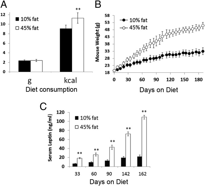
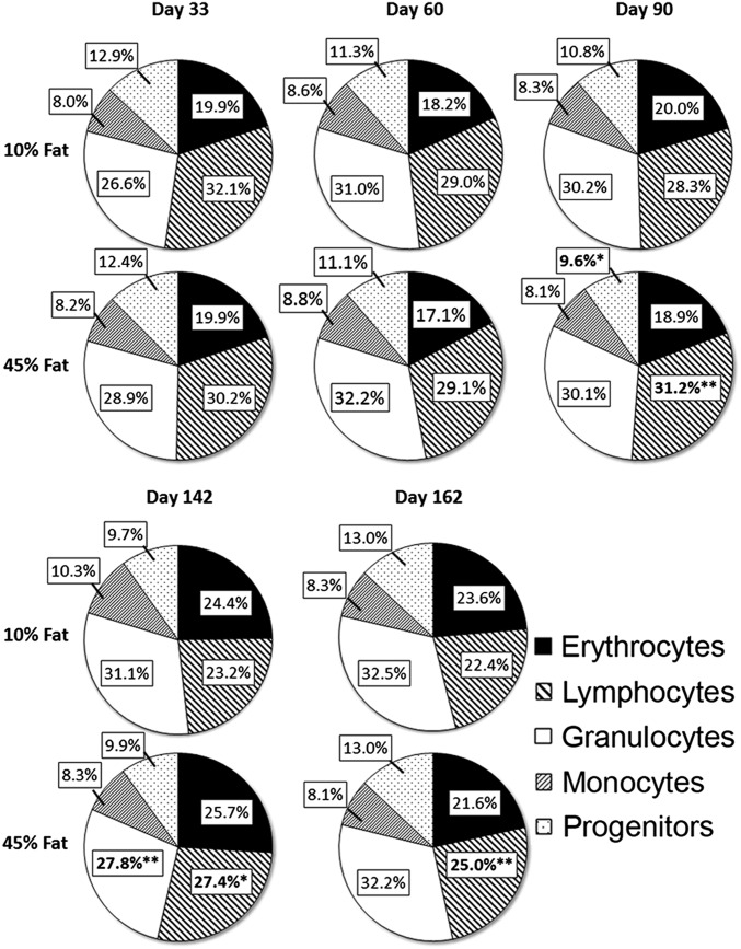
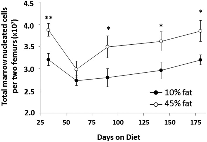
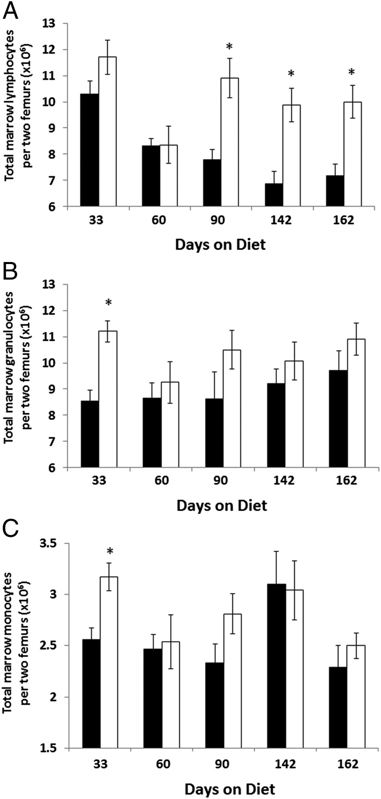
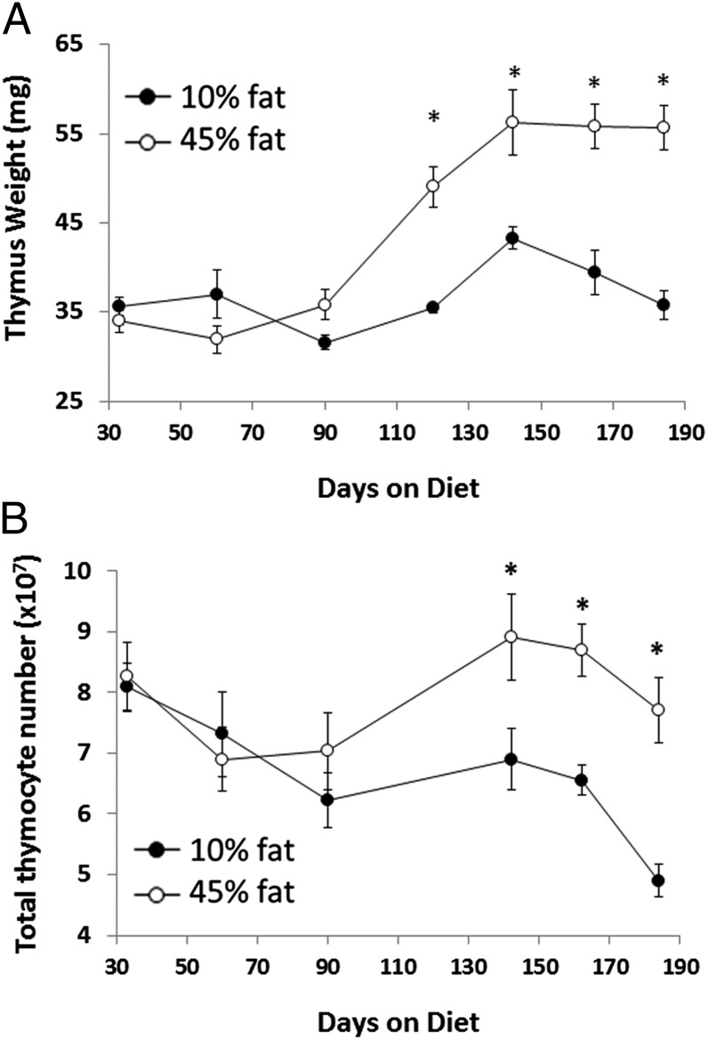
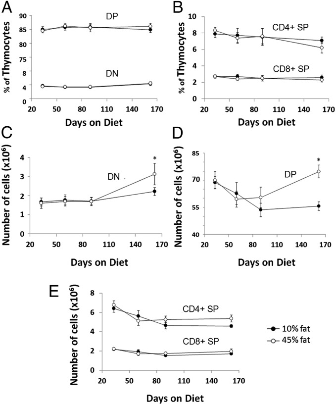
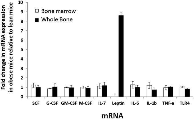
A rodent model of diet-induced obesity revealed that obesity significantly altered hematopoietic and lymphopoietic functions in the bone marrow and thymus. C57BL/6 mice were fed a mixed high-fat diet (HFD) of 45% fat or 10% fat diet (lean controls) for 180 d. A sustained increase in the numbers of cells found in bone marrow and thymus of HFD mice occurred from day 90 to day 180. However, with the exception of a 10-18% increase in the proportion of lymphocytes, the composition of monocytes, granulocytes, erythrocytes, and mixed progenitor lineages remained normal in the marrow. Likewise, thymuses of HFD mice increased 30-50% in size compared with controls, with analogous increases in thymocyte numbers. The overall thymus cellular composition remained normal. Although increased blood and lymphatic volume in obese mice would play a role in increased hematopoiesis, there were large and disproportionate increases in blood leukocytes of HFD mice, indicating that homeostasis was not maintained. Leptin, which promotes lymphopoiesis and myelopoiesis, reached 100 ng/mL in sera from HFD mice. Moreover, a three- to sixfold increase in adipocytes in marrow resulted in spiked leptin mRNA expression in bones of HFD mice compared with lean controls. Other cytokines and growth factors did not show any increases in obese marrow. The substantial increase in lymphopoietic and hematopoietic processes in HFD mice indicates that the primary tissues are another facet of the immune system dysregulated by obesity, which was perhaps fostered by higher amounts of leptin in marrow and serum.


Conflict of interest statement

The authors declare no conflict of interest.

Figures

Fig. 1.
Fig. 1.
Characteristics of mice fed HFD. Mice fed a 45% mixed fat diet (white bars) consumed similar amounts of food in grams as mice fed a 10% fat diet (black bars) but ingested significantly more calories (A). Data shown are average food consumption per day for the entire 180-d experiment. (B) Mice fed a 45% fat diet (○) gained significantly more weight than mice fed a 10% fat diet (●). (C) Serum leptin in mice fed a 45% fat diet (white bars) was significantly elevated compared with mice fed a 10% fat diet (black bars; n = 7–8 mice per group). **P < 0.01.
Fig. 2.
Fig. 2.
Distribution of bone marrow hematopoietic cell subpopulations in mice fed 10% or 45% fat diet. Bone marrow cells were labeled with anti-CD31 (clone ER-MP12) and anti–Ly-6C (clone ER-MP20), with cell subpopulations determined as described in Fig. S2, including specific markers to confirm the identity and size of each marrow subpopulation. Data show the percent of total nucleated marrow cells for each cell type at various times after the start of diet. n = 7–8 mice per group. *P < 0.05, **P < 0.01.
Fig. 3.
Fig. 3.
Increased numbers of nucleated cells in bone marrow of obese mice. Mice fed the 45% fat diet (○) showed increased bone marrow cell numbers compared with mice fed the 10% fat diet (●). Data shown are the total numbers of nucleated marrow cells per two femurs. n = 7–8 mice per group. *P < 0.05, **P < 0.01.
Fig. 4.
Fig. 4.
Total numbers of different bone marrow cell subclasses. Mice fed a 45% fat diet (white bars) had increased numbers of lymphocytes (CD31+/Ly-6C/B220+) in their bone marrow compared with mice fed a 10% fat diet (black bars) starting at day 90 on diet (A). Total numbers of (B) marrow granulocytes (CD31/Ly-6C+/Ly-6G+/Gr-1+) and (C) monocytes (CD31+/Ly-6Chi/Ly-6G/Gr-1+) were only modestly affected. Data represent total numbers of each marrow cell type in two femurs. n = 7–8 mice per group. *P < 0.05.
Fig. 5.
Fig. 5.
Thymus size and cell number were elevated in HFD mice. Mice fed a 45% fat diet (○) had increased thymus size (A) and total thymocyte number (B) beyond 120 d on diet compared with mice fed a 10% fat diet (●). n = 7–8 mice per group. *P < 0.05.
Fig. 6.
Fig. 6.
Thymus cellular composition and total numbers in HFD mice. Thymuses from mice fed a 45% fat diet (○) were analyzed by flow cytometry for thymus cell subpopulations and compared with mice fed a 10% fat diet (●). The composition of thymocyte subclasses as defined by CD4 and CD8 expression was not significantly altered by obesity (A and B). When total numbers of thymocytes were assessed, significant increases in (C) DN (CD4/CD8) and (D) DP (CD4+/CD8+) subtypes were observed at day 162 on diet, when thymus size was significantly larger in obese mice compared with controls. No significant alterations were seen in total numbers of mature single positive CD4+/CD8 and CD4/CD8+ thymocytes (E). n = 7–8 mice per group. *P < 0.05.
Fig. 7.
Fig. 7.
Analysis of expression of various genes in isolated bone marrow cells that would not contain adipocytes because of processing method and whole bone that would contain adipocytes from HFD mice. Total RNA was isolated bone marrow cells (white bars) and whole bone (black bars) and subjected to RT-PCR for relative quantitation of mRNA expression. Data shown represent expression of each mRNA in obese mice relative to expression in control lean mice. Data represent the average of two experiments for bone marrow and whole bone. For marrow, cells from days 142 and 165 on diet were used. For whole bone, femurs from days 157 and 165 were used. n = 8 per group at each time point. –, not detected.
Fig. 8.
Fig. 8.
Quantification of adipocytes in bone marrow of HFD mice. Femurs from mice fed 10% or 45% fat diet were sectioned and stained with H&E for visualization of adipocytes. Adipocyte number was determined by counting the number of adipocytes per femur section using a light microscope (two sections per animal). Each circle represents one mouse, and black bars indicate mean for that group. n = 7–8 mice per group. *P < 0.05.

References

    1. Hardy RR, et al. B-cell commitment, development and selection. Immunol Rev. 2000;175:23–32. - PubMed
    1. Rolink AG, Schaniel C, Andersson J, Melchers F. Selection events operating at various stages in B cell development. Curr Opin Immunol. 2001;13:202–207. - PubMed
    1. Fraker PJ, King LE. Reprogramming of the immune system during zinc deficiency. Annu Rev Nutr. 2004;24:277–298. - PubMed
    1. Lodish H, Flygare J, Chou S. From stem cell to erythroblast: Regulation of red cell production at multiple levels by multiple hormones. IUBMB Life. 2010;62:492–496. - PMC - PubMed
    1. Ueda Y, Kondo M, Kelsoe G. Inflammation and the reciprocal production of granulocytes and lymphocytes in bone marrow. J Exp Med. 2005;201:1771–1780. - PMC - PubMed

Publication types

MeSH terms