Comparative Study
. 2013 Jun;35(3):747-62.
doi: 10.1007/s11357-012-9407-9. Epub 2012 Apr 27.

MicroRNA-22 increases senescence and activates cardiac fibroblasts in the aging heart

Affiliations
Comparative Study

MicroRNA-22 increases senescence and activates cardiac fibroblasts in the aging heart

Virginija Jazbutyte et al. Age (Dordr). 2013 Jun.

Abstract

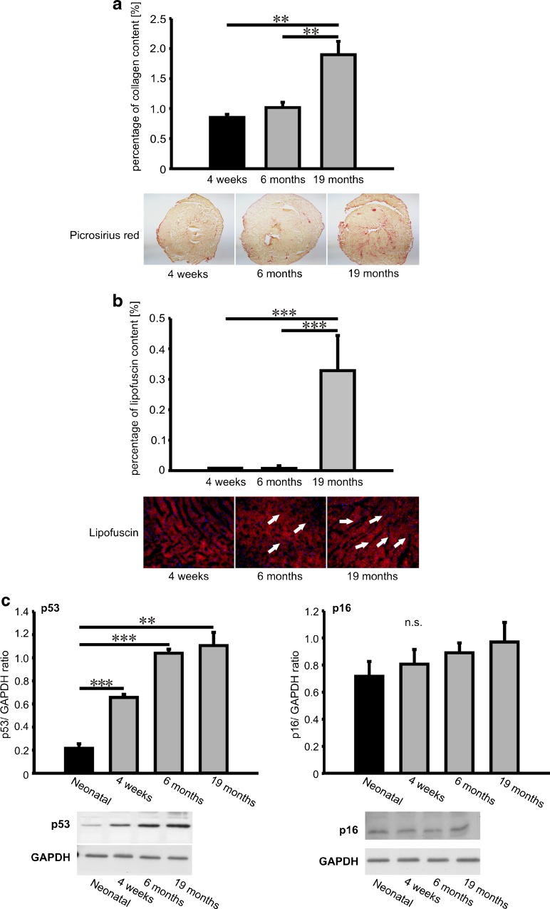
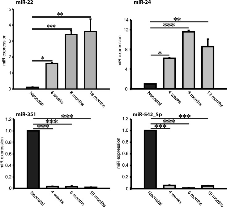
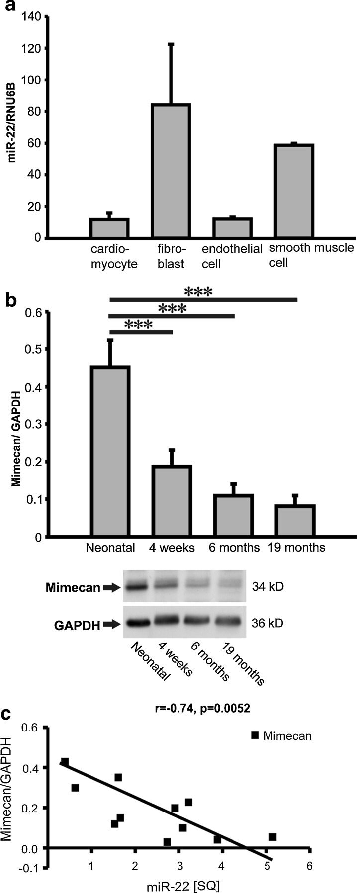
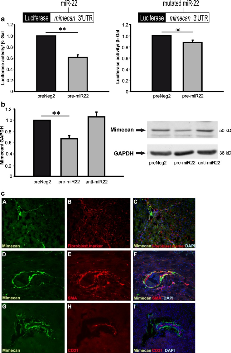
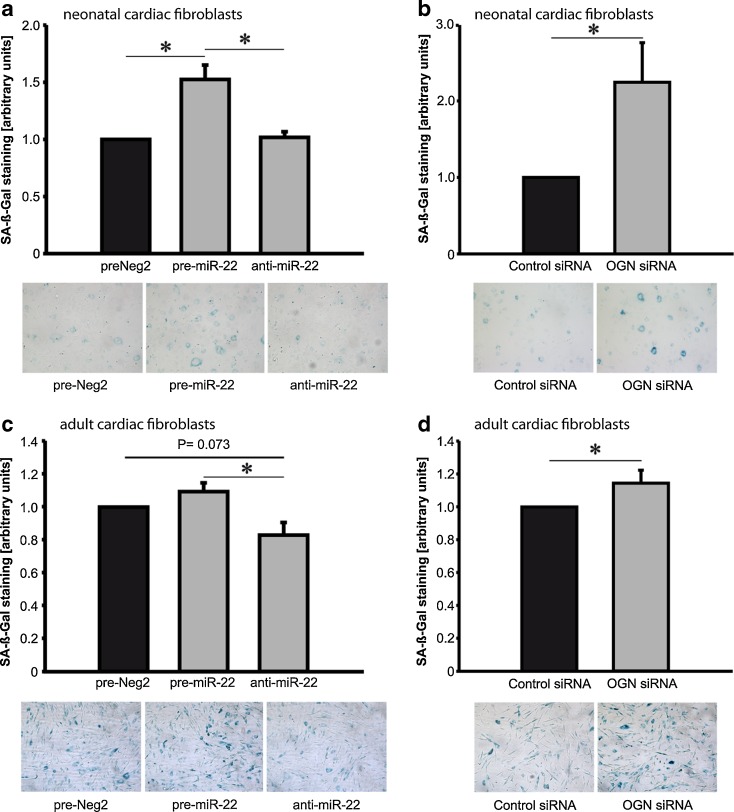
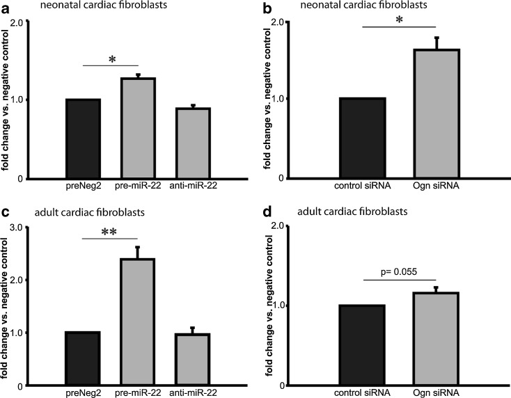
MicroRNAs (miRs) are small non- coding RNA molecules controlling a plethora of biological processes such as development, cellular survival and senescence. We here determined miRs differentially regulated during cardiac postnatal development and aging. Cardiac function, morphology and miR expression profiles were determined in neonatal, 4 weeks, 6 months and 19 months old normotensive male healthy C57/Bl6N mice. MiR-22 was most prominently upregulated during cardiac aging. Cardiac expression of its bioinformatically predicted target mimecan (osteoglycin, OGN) was gradually decreased with advanced age. Luciferase reporter assays validated mimecan as a bona fide miR-22 target. Both, miR-22 and its target mimecan were co- expressed in cardiac fibroblasts and smooth muscle cells. Functionally, miR-22 overexpression induced cellular senescence and promoted migratory activity of cardiac fibroblasts. Small interference RNA-mediated silencing of mimecan in cardiac fibroblasts mimicked the miR-22-mediated effects. Rescue experiments revealed that the effects of miR-22 on cardiac fibroblasts were only partially mediated by mimecan. In conclusion, miR-22 upregulation in the aging heart contributed at least partly to accelerated cardiac fibroblast senescence and increased migratory activity. Our results suggest an involvement of miR-22 in age-associated cardiac changes, such as cardiac fibrosis.


Figures

Fig. 1
Fig. 1
Cardiac fibrosis and aging (lipofuscin deposition, p53 and p16 expression). Cardiac fibrosis was visualized by picrosirius red (PSR) staining where collagen fibers are red whereas myocardium is stained in yellow. The degree of cardiac fibrosis is expressed as percentage of collagen signal versus total signal intensity (chart in Fig. 1a ). Representative PSR stainings are shown in the middle panel. Deposition of autofluorescent lipofuscin particles was observed in 4 % PFA fixated cryosections using fluorescence filter at 594 nm wavelength range (lower panel). Quantification data of lipofuscin signal intensity in cardiac sections are expressed as percentage of lipofuscin signal versus total signal intensity (chart, Fig. 1b). c Protein expression of senescence-associated p53 and p16 in cardiac tissue. Data in the chart are expressed as mean ± SEM (n = 4 tissues/sections/ group); **, p < 0.01; ***, p < 0.001
Fig. 2
Fig. 2
Postnatally differentially regulated microRNAs. Differentially expressed microRNAs in hearts from neonatal, 4 months, 6 months and 19 months old male Bl6 mice. MiR-22 and miR-24 were strongly upregulated whereas miR-351 and miR-542-5p were significantly downregulated in postnatal compared to neonatal hearts. Data are expressed as mean ± SEM (n = 8/ group). *, p < 0.05; **, p < 0.01; ***, p < 0.001
Fig. 3
Fig. 3
MiR-22 and mimecan expression in the aging heart. a MiR-22 expression in rat cardiac myocytes, human cardiac fibroblasts, umbilical cord endothelial cells and smooth muscle cells showed miR-22 enrichment in cardiac fibroblasts and smooth muscle cells (n = 3- 4/ group). b Protein expression levels of the miR-22 predicted target mimecan in hearts from aging mice. c Negative correlation between miR-22 and mimecan in the mouse hearts depicted by regression analysis with a Pearson’s correlation coefficient of r = -0.74 (p = 0.0052). Data are expressed as mean SQ ± SEM. *, p < 0.05; **, p < 0.01; ***, p < 0.001
Fig. 4
Fig. 4
Mimecan is regulated by miR-22 and localized in the mouse heart. a Luciferase reporter vector carrying either wild- type (left panel) or mutant human mimecan 3’- UTR (right panel) was co-transfected with control miR (preNeg2) or miR-22 precursor (pre-miR22). Luciferase activity results were normalized versus beta galactosidase activity in the same cells. b MiR-22 overexpression resulted in significant mimecan protein downregulation in cardiac fibroblasts, whereas inhibition of endogenous miRNA by sequence- specific antagonist did not influence mimecan expression. Representative mimecan western blots are shown in b, right panel. c Immunofluorescent staining of mimecan protein expression (green) in the murine heart. Mimecan was partially co- localized with cardiac fibroblasts (red) (panels a-c) and smooth muscle cells (red) (smooth muscle actin (SMA) staining) (panels d-f) but not with endothelial cells (red) (marker CD31) (g-i). Cell nuclei were stained with DAPI. The pictures were taken at 200x magnification. Data are expressed as mean ± SEM (n = 3-5/ group). **, p < 0.01
Fig. 5
Fig. 5
Effects of miR-22 on cellular senescence. Neonatal rat and adult human cardiac fibroblasts were transfected with miR-22 precursor (pre-miR-22), miR-22 antagonist (antimiR-22) or control miR (preNeg2) (a, c). To determine enzymatic activity of senescence-associated beta galactosidase (SA-β- Gal), cells were fixed and incubated with β- Gal substrate. Blue color intensity was analyzed and data was expressed as ratio between treatment and control group. MiR-22 overexpression significantly induced premature senescence in neonatal rat cardiac fibroblasts (a). Mimecan (osteoglycin, OGN) silencing using sequence- specific siRNA induced cellular senescence in neonatal fibroblasts mimicking the effects of miR-22 overexpression (b). Adult cardiac fibroblasts expressed high endogenous levels of miR-22, thus overexpression of this microRNA resulted in marginal increase in cellular senescence, whereas miR-22 inhibition reduced it (c). Mimecan (OGN) silencing increased cellular senescence in adult cardiac fibroblasts (d). Data are expressed as mean ± SEM (n = 4-5/ group). *, p < 0.05
Fig. 6
Fig. 6
Chemotactic migratory capacity of cardiac fibroblasts is mediated by miR-22. Neonatal rat and adult human cardiac fibroblasts were transfected with miR-22 precursor, antagonist and control miR (preNeg 2). Chemotactic migration assay was performed using modified Boyden chamber approach where cells migrated through semi- permeable membrane towards 20 % FCS. Migrated cells were stained with DAPI and counted. The mean number of migrated neonatal fibroblasts transfected with control miRNA was set to 1 and the ratios between pre-miR-22 and anti-miR-22 versus group and control are depicted in panel a. MiR-22- induced chemotactic activity of fibroblasts could be mimicked by mimecan (Ogn) knockdown in neonatal fibroblasts (b). Similarly, ectopic expression of miR-22 in adult fibroblasts also resulted in significant increase in chemotaxis (c) and these effects could be reproduced by mimecan (Ogn) knockdown (panel d, p = 0.055). Data are expressed as mean ± SEM (n = 4-5/ group). *, p < 0.05

References

    1. Bauersachs J, Thum T. Biogenesis and regulation of cardiovascular microRNAs. Circ Res. 2011;109(3):334–347. doi: 10.1161/CIRCRESAHA.110.228676. - DOI - PubMed
    1. Bentz H, Nathan RM, Rosen DM, Armstrong RM, Thompson AY, Segarini PR, Mathews MC, Dasch JR, Piez KA, Seyedin SM. Purification and characterization of a unique osteoinductive factor from bovine bone. J Biol Chem. 1989;264(34):20805–20810. - PubMed
    1. Blakaj A, Lin H. Piecing together the mosaic of early mammalian development through microRNAs. J Biol Chem. 2008;283(15):9505–9508. doi: 10.1074/jbc.R800002200. - DOI - PMC - PubMed
    1. Brodersen P, Voinnet O. Revisiting the principles of microRNA target recognition and mode of action. Nat Rev. 2009;10(2):141–148. doi: 10.1038/nrm2619. - DOI - PubMed
    1. Coolen M, Bally-Cuif L. MicroRNAs in brain development and physiology. Curr Opin Neurobiol. 2009;19(5):461–470. doi: 10.1016/j.conb.2009.09.006. - DOI - PubMed

Publication types