. 2012 Jul 1;87(1):11, 1-12.
doi: 10.1095/biolreprod.112.099390. Print 2012 Jul.

Zinc maintains prophase I arrest in mouse oocytes through regulation of the MOS-MAPK pathway

Affiliations

Zinc maintains prophase I arrest in mouse oocytes through regulation of the MOS-MAPK pathway

Betty Y Kong et al. Biol Reprod. .

Abstract

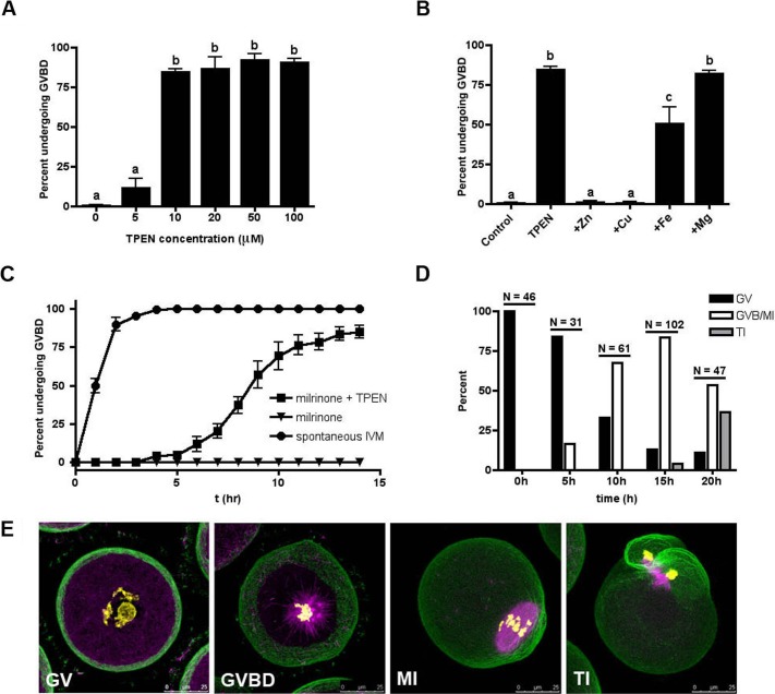
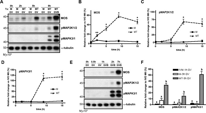
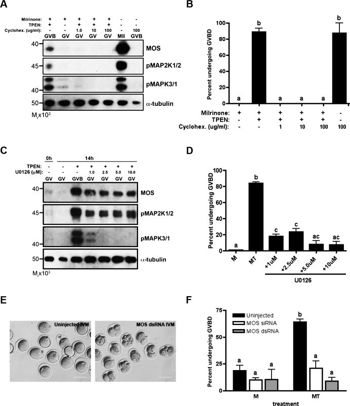
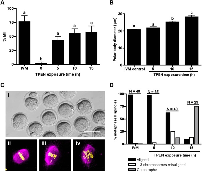
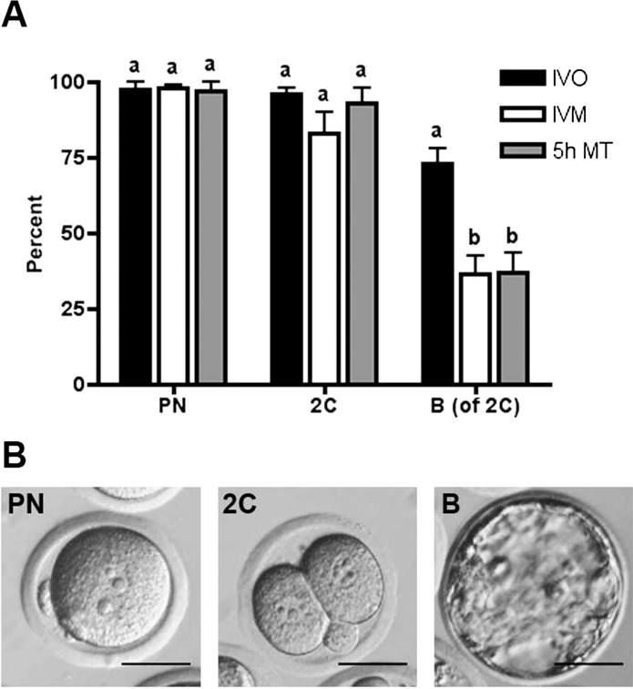
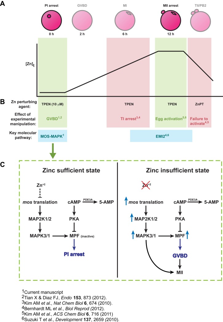
Meiosis in mammalian females is marked by two arrest points, at prophase I and metaphase II, which must be tightly regulated in order to produce a haploid gamete at the time of fertilization. The transition metal zinc has emerged as a necessary and dynamic regulator of the establishment, maintenance, and exit from metaphase II arrest, but the roles of zinc during prophase I arrest are largely unknown. In this study, we investigate the mechanisms of zinc regulation during the first meiotic arrest. Disrupting zinc availability in the prophase I arrested oocyte by treatment with the heavy metal chelator N,N,N',N'-tetrakis-(2-pyridylmethyl)-ethylenediamine (TPEN) causes meiotic resumption even in the presence of pharmacological inhibitors of meiosis. We further show that the MOS-MAPK pathway mediates zinc-dependent prophase I arrest, as the pathway prematurely activates during TPEN-induced meiotic resumption. Conversely, inhibition of the MOS-MAPK pathway maintains prophase I arrest. While prolonged zinc insufficiency ultimately results in telophase I arrest, early and transient exposure of oocytes to TPEN is sufficient to induce meiotic resumption and bypass the telophase I block, allowing the formation of developmentally competent eggs upon parthenogenetic activation. These results establish zinc as a crucial regulator of meiosis throughout the entirety of oocyte maturation, including the maintenance of and release from the first and second meiotic arrest points.


Figures

FIG. 1.
FIG. 1.
Zinc insufficiency induces meiotic resumption with prolonged meiosis I kinetics. A) The incidence of GVBD was scored following 14-h culture of DOs in the presence of milrinone and increasing concentrations of TPEN. B) To determine the zinc specificity of TPEN-mediated GVBD, medium containing milrinone and 10 μM TPEN was supplemented with equimolar concentrations of zinc, iron, copper, or magnesium. Oocytes were scored for incidence of GVBD at 14 h. C) The kinetics of GVBD were determined in DOs cultured for 14 h in medium without milrinone (i.e., in vitro matured; circles), medium containing milrinone (triangles), or medium containing milrinone and TPEN (squares). D) The specific meiotic stage of oocytes cultured in the presence of milrinone and TPEN was quantified by analysis of α-tubulin (magenta), F-actin (green), and chromatin (yellow) immunostaining. Oocytes were scored as GV, GVBD/MI, or TI according to the status of the cytoskeleton and the chromosome configuration. “N” represents the total number of oocytes examined at each time point. E) Representative confocal images of each category of oocytes are shown as Z stack projections of sections containing the spindle and/or chromatin. Bars = 25 μm. Experiments in AC were conducted at least three times and the results represent the mean ± SEM. Between 30 and 60 oocytes were examined per group per experimental repeat. In A and B, statistical differences in the incidence of GVBD were calculated according to one-way ANOVA with Bonferroni post hoc test and labeled as indicated (P < 0.001).
FIG. 2.
FIG. 2.
TPEN-treated oocytes prematurely accumulate MOS and activate the MOS-MAPK pathway. A) Lysates from oocytes cultured in milrinone alone (M) or milrinone and TPEN (MT) were collected at the indicated time points and immunoblotted for MOS, phospho-MAP2K1/2, and phospho-MAPK3/1. BD) Protein levels of MOS (B), phospho-MAP2K1/2 (C), and phospho-MAPK3/1 (D) ± SEM were quantified by densitometric analysis of at least three experiments normalized to α-tubulin. *P < 0.05, **P < 0.005 according to Student t-test, comparing TPEN-treated (MT) to untreated (M) oocytes at each time point. E) Lysates from oocytes were collected at the indicated time points during IVM for immunoblotting. Note that, as compared to TPEN-induced GVBD, protein levels of MOS, phospho-MAP2K1/2, and phospho-MAPK3/1 in in vitro matured oocytes do not increase until after GVBD has occurred. F) Blots similar to those represented in A and E were analyzed to compare the protein levels of GV intact oocytes collected after 1-h culture in IVM medium (black bars), 9-h culture in milrinone medium (white bars), and 9-h culture in milrinone-TPEN medium (grey bars). Analysis was performed by densitometry of at least three experiments and normalized to α-tubulin. For each protein, groups with uncommon letters indicate statistical differences in the protein level (P < 0.05). All immunoblots contain 20 oocytes per lane.
FIG. 3.
FIG. 3.
TPEN-induced meiotic resumption is dependent on activation of the MOS-MAPK pathway. Global protein synthesis was inhibited by addition of cycloheximide to culture (A and B). The MOS-MAPK was specifically inhibited by treatment with U0126 (C and D), or microinjection of Mos hairpin dsRNA or siRNA (E and F). A and C) Lysates obtained from TPEN-treated and in vitro-matured oocytes cultured for 14 h in the presence of cycloheximide (A) or U0126 (C) were immunoblotted for MOS, phospho-MAP2K1/2, and phospho-MAPK3/1 expression. Each lane contains 20 oocytes. B and D) The incidence of GVBD after cycloheximide (B) or U0126 (D) treatment was scored by light microscopy. E) Oocytes were injected with MOS hairpin dsRNA, held for 10–14 h, and in vitro matured. Functionality of dsRNA injection was determined by visualization of parthenogenetic activation after 36-h culture. Bars = 80 μm. F) Oocytes injected with MOS hairpin dsRNA or MOS siRNA were held for 10–14 h and either maintained in milrinone or transferred to medium containing milrinone and TPEN for an additional 14 h. The incidence of GVBD was scored. In B, D, and F, experiments were performed at least three times and the results represent the mean ± SEM. Between 30 and 60 oocytes were analyzed per group per experimental repeat. Groups with uncommon letters indicate statistical differences in the percentage of oocytes undergoing GVBD (P < 0.001). M, milrinone only; MT, milrinone and TPEN.
FIG. 4.
FIG. 4.
Transient TPEN exposure permits meiotic progression to MII. A) Oocytes were cultured in milrinone-TPEN medium for the indicated times and transferred to medium without TPEN for a total of 24 h. Progression to MII was assessed by scoring for the presence of a PB. The experiment was repeated three times and the results represent the mean ± SEM. Approximately 30 oocytes were used per group per experiment. B) PB diameter was measured by taking the average of two perpendicular lengths and compared among treatment groups. Results represent the mean ± SEM. C, i) A representative image of MII eggs obtained from TPEN exposure followed by culture in milrinone. C, ii–iv) To determine the quality of the MII spindle, eggs that had a visible PB by light microscopy were fixed and stained for α-tubulin (magenta) and chromatin (yellow). Chromosomes on the metaphase plate were categorized as aligned (i), one to three misaligned (ii), or catastrophe (iii). Representative confocal images of each category are shown as Z stack projections. Bar in i = 80 μm; bars in iiiv = 10 μm. D) Chromosome alignment was scored according to the categories described in C. “N” represents the total number of eggs examined for each culture condition. Only spindles oriented approximately parallel to the plane of the Z stacks were scored. In A and B, groups with uncommon letters indicate statistical differences (P < 0.01).
FIG. 5.
FIG. 5.
MII eggs derived after TPEN exposure are developmentally competent. MII eggs obtained after transient TPEN exposure followed by a 14-h culture in IVM medium were parthenogenetically activated. A) Following parthenogenetic activation, the preimplantation developmental progression of eggs derived from TPEN-treatment (5h MT; gray bars), in vivo ovulation (black bars), and IVM (white bars) was scored. Specifically, the progression to pronuclear (PN), two-cell (2C), and blastocyst stage (B) were analyzed by light microscopy, and representative images are shown in B. The percentages of PN and 2C were calculated based on total number of MII eggs. The percentage of blastocysts was calculated based on number of 2C embryos. The experiment was performed three times and results represent the mean ± SEM. Between 20 and 40 eggs were used per group per experiment. For each embryo stage, groups with uncommon letters indicate statistical differences (P < 0.05). Bars = 40 μm.
FIG. 6.
FIG. 6.
A) Total cellular zinc ([Zn]t) increases by 50% during meiotic maturation from the PI arrested oocyte to the MII arrested egg, and then drops upon fertilization in events described as zinc sparks. B) Perturbation of zinc availability using the heavy metal chelator TPEN or the zinc ionophore ZnPT at critical windows of meiotic maturation and fertilization interferes with specific zinc-dependent pathways, resulting in premature arrest or progression through the maturation pathway. Decreasing zinc availability using TPEN at the two crucial arrest points, namely PI and MII, results in premature meiotic progression (green shading). Oocytes that undergo GVBD due to zinc insufficiency have the capacity to develop to MII eggs upon return to zinc-replete medium and blastocysts upon egg activation. This effect of zinc insufficiency in causing meiotic resumption from PI arrest is mediated via the MOS-MAPK pathway. As described in Supplemental Figure S1, experimental manipulation of zinc levels before or after MII arrest, when zinc is actively being acquired or released, respectively, results in meiotic arrest (red shading). C) Model showing proposed action of a zinc-mediated pathway during the first meiotic arrest at PI. In a zinc-sufficient state, the MOS-MAPK pathway is maintained in a quiescent state, MPF activity remains low, and the oocyte remains arrested at the PI stage. Zinc insufficiency, as induced by chelation, relieves the inhibition on the MOS-MAPK pathway, allowing meiotic resumption, GVBD, and progression toward MII.

References

    1. Tsafriri A. Mammalian oocyte maturation: model systems and their physiological relevance. Adv Exp Med Biol 1979; 112: 269 281 - PubMed
    1. Josefsberg LB, Galiani D, Lazar S, Kaufman O, Seger R, Dekel N. Maturation-promoting factor governs mitogen-activated protein kinase activation and interphase suppression during meiosis of rat oocytes. Biol Reprod 2003; 68: 1282 1290 - PubMed
    1. Conti M, Andersen CB, Richard F, Mehats C, Chun SY, Horner K, Jin C, Tsafriri A. Role of cyclic nucleotide signaling in oocyte maturation. Mol Cell Endocrinol 2002; 187: 153 159 - PubMed
    1. Paules RS, Buccione R, Moschel RC, Vande Woude GF, Eppig JJ. Mouse Mos protooncogene product is present and functions during oogenesis. Proc Natl Acad Sci U S A 1989; 86: 5395 5399 - PMC - PubMed
    1. Shibuya EK, Ruderman JV. Mos induces the in vitro activation of mitogen-activated protein kinases in lysates of frog oocytes and mammalian somatic cells. Mol Biol Cell 1993; 4: 781 790 - PMC - PubMed

Publication types

MeSH terms