. 2012 May 25;110(11):1465-73.
doi: 10.1161/CIRCRESAHA.112.269035. Epub 2012 Apr 26.

MicroRNA-mediated in vitro and in vivo direct reprogramming of cardiac fibroblasts to cardiomyocytes

Affiliations

MicroRNA-mediated in vitro and in vivo direct reprogramming of cardiac fibroblasts to cardiomyocytes

Tilanthi M Jayawardena et al. Circ Res. .

Abstract

Rationale: Repopulation of the injured heart with new, functional cardiomyocytes remains a daunting challenge for cardiac regenerative medicine. An ideal therapeutic approach would involve an effective method at achieving direct conversion of injured areas to functional tissue in situ.

Objective: The aim of this study was to develop a strategy that identified and evaluated the potential of specific micro (mi)RNAs capable of inducing reprogramming of cardiac fibroblasts directly to cardiomyocytes in vitro and in vivo.

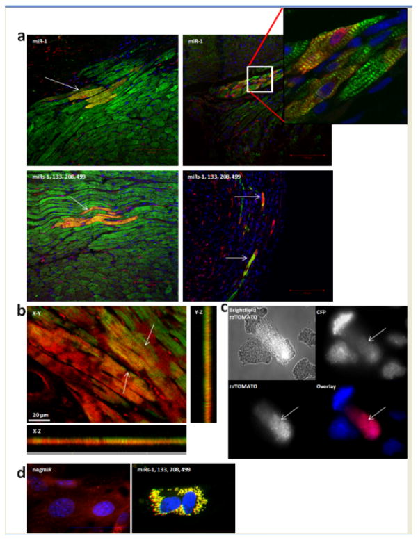
Methods and results: Using a combinatorial strategy, we identified a combination of miRNAs 1, 133, 208, and 499 capable of inducing direct cellular reprogramming of fibroblasts to cardiomyocyte-like cells in vitro. Detailed studies of the reprogrammed cells demonstrated that a single transient transfection of the miRNAs can direct a switch in cell fate as documented by expression of mature cardiomyocyte markers, sarcomeric organization, and exhibition of spontaneous calcium flux characteristic of a cardiomyocyte-like phenotype. Interestingly, we also found that miRNA-mediated reprogramming was enhanced 10-fold on JAK inhibitor I treatment. Importantly, administration of miRNAs into ischemic mouse myocardium resulted in evidence of direct conversion of cardiac fibroblasts to cardiomyocytes in situ. Genetic tracing analysis using Fsp1Cre-traced fibroblasts from both cardiac and noncardiac cell sources strongly suggests that induced cells are most likely of fibroblastic origin.

Conclusions: The findings from this study provide proof-of-concept that miRNAs have the capability of directly converting fibroblasts to a cardiomyocyte-like phenotype in vitro. Also of significance is that this is the first report of direct cardiac reprogramming in vivo. Our approach may have broad and important implications for therapeutic tissue regeneration in general.


Figures

Figure 1
Figure 1. Introduction of microRNA(s) into cardiac fibroblasts induces the expression of cardiac myocyte-specific markers
a, Cumulative gene expression data from miRNA-transfected adult cardiac fibroblasts are illustrated graphically in heat map form. These results depict a major shift in fibroblastic (Ddr2 & Vim) to early cardiomyocyte (Isl1, Nkx2.5, Gata4, Hand2, Mef2c, and late cardiac myocyte (Tnni3, Cx43) marker expression between 3 and 6 days post-transfection. Fold change in normalized gene expression for each gene is shown on a green to black to red scale (Minimum(Green) – Maximum(Red). Detailed color legends describing quantitative fold change data for each gene are represented in Online Figure XX. b, Representative bar graphs showing RNA expression profiles of Gata4 and Mef2c in neonatal cardiac fibroblasts at 3 days post-transfection. Highlighted are top miRNA combinations 1, 133, 206 and 1, 133, 208. All miRNA combinations represented by light grey bars. Dark grey bar represents averaged controls; untransfected, mock and non-targeting miRNA (negmiR). Results presented as mean ± SEM. c, Representative scatter plot showing the geometric mean of normalized expression of Gata4, Mef2c, and Tbx5 at 3 days post-transfection. Highlighted are: miRs-1, 133, 208 (red); and miRs-1, 133, 206, (purple); mock, negmiR, and untransfected controls (green); remaining miRNA combinations (black). d, α-ACTININ (in green) immunostaining of DAPI-postive (blue) neonatal cardiac fibroblasts 1 week following transfection with miR-1; miRs-1, 133, 206; miRs-1, 133, 208; and miRs-133, 206, 208. Scale bar, 100μm. e, TNNI3 (in green) immunostaining of DAPI-positive (blue) neonatal cardiac fibroblasts 1 week following transfection with miRs-1, 133, 208. Zoomed in area highlights the presence of prominent striations in TNNI3+ cells. f, TNNI3 (in green) immunostaining of DAPI-postive (blue) neonatal cardiac fibroblasts isolated from Fsp1Cre/tdTOMATO (red) mice, 4 weeks following transfection with miRs-1, 138, 208. Scale bar, 100μm.
Figure 2
Figure 2. Cardiac induction is enhanced by the addition of miR-499 and /or small molecule inhibitor of JAK Inhibitor I
a, Summary from FACS analyses data of miRNA-transfected neonatal cardiac fibroblasts. Cardiac fibroblasts were isolated from αMHC-CFP neonatal transgenic mice and induced CFP+ population was analyzed and sorted by FACS 7 days after transfection. In samples involving JAK inhibitor I treatment, miRNA-transfected cells were treated with the small molecule 48 hours post-transfection and then sorted by FACS. Provided is the range of %CFP induction for each miRNA combination +/− JAK inhibitor I for several experimental rounds (n=4–8). b, RNA expression of cardiac myocyte-specific markers Mef2c, Tnni3, and Myh6 in CFP+ cells sorted from neonatal cardiac fibroblasts 1 week following transfection with miR-1 and miRs-1, 133, 208, 499. c, Representative FACS analyses demonstrating the induction of αMHC-driven-CFP+ cell population in miRNA-transfected neonatal cardiac fibroblasts with and without treatment of JAK inhibitor I, 1 week post-transfection. FACS traces are distinguished as follows: untransfected cells (green), negmiR-transfected cells (red) and miRNA-transfected cells (light blue). For b, results presented as mean ± SEM. * P<0.05, one way ANOVA with Bonferroni correction. Stated p value is versus non-targeting miRNA control. d, RNA expression levels of cardiac ion channels, Cacna1c, Scn5a, and Kcnj2, in CFP+ cells sorted from JAK inhibitor I-treated neonatal cardiac fibroblasts, 1 week following transfection with miR-1 and miRs-1, 133, 208, 499. Both untransfected and negmiR controls were also treated with the JAK inhibitor I. For f, results are presented as mean ± SEM. * P<0.05, one way ANOVA with Bonferroni correction. Stated p value is versus negmiR control.
Figure 3
Figure 3. miRNA-transfected cardiac fibroblasts exhibit spontaneous calcium oscillations and calcium transients in response to depolarization
a, Representative examples of spontaneous calcium oscillations in CFP+ cells 14 days post-FACS from JAK inhibitor I-treated cardiac fibroblasts transfected with miRNAs, or negmir and in neonatal cardiac myocytes. b, Average oscillation frequencies in miR-1 (n=17) and miR-1, 133, 208, 499 (n=40) transfected cardiac fibroblasts at 14 days post-FACS. Controls are untransfected (n=1) and negmir-transfected (n=1) cardiac fibroblasts and neonatal cardiomyocytes (n=88). All miRNA-transfected and control cardiac fibroblasts were subjected to JAK inhibitor I treatment. c, Total number of oscillating cells in miRNA-induced, JAK inhibitor I-treated CFP+ and control cell populations at 14 days post-FACS (expressed as a percentage of the number observed in miR-1, 133, 208, 499-induced CFP+ population). d, Representative example of a calcium response to depolarization with high [K+] (60 mM) in miR-1-induced CFP+ cells at 14 days post-FACS. High [K+] (2 min exposure) induced the onset of calcium oscillations that persisted following reintroduction of low [K+] Ringer. e, Percentage of miR-1 (n=19), miR-1, 133, 208, 499 (n=13), and negmir (n=11) transfected and untransfected (n=16) cardiac fibroblasts that exhibited a calcium response to depolarization with high [K+]. These data were collected from two independent experimental rounds, with at least 8 fields of cells sampled from each. All miRNA-transfected and control cells were subjected to JAK inhibitor I treatment.
Figure 4
Figure 4. MicroRNA-injected endogenous cardiac fibroblasts are reprogrammed in vivo following myocardial injury
a, Fsp1Cre/tdTOMATO transgenic mice were injected with miRNAs intramyocardially at the time of permanent ligation of the left ascending coronary artery (LAD). Four weeks later, hearts were harvested, fixed, and immunostained with antibodies against tdTOMATO (red) and cardiac troponin T (TNNT2) (green). Nuclear staining with DAPI(blue). For split channel images, please refer to Online Figure XI. Large, double-positive cells (red and green) with prominent striations (highlighted with arrows), often organized as part of clusters, were taken as evidence of reprogrammed cardiac fibroblasts. Shown here are representative images from infarct and peri-infarct areas of miR-1 and miRs-1, 133, 208, 499-(miR combo) injected hearts. Scale bar, 100 μm. Zoomed in area highlights a representative example of the presence of prominent striations in tdTOMATO+ TNNT2+ cells from miRNA-injected hearts. b, Zoom of selected confocal microscopy image from the heart of a miRNA-injected heart from a Fsp1Cre-tdTOMATO animal; orthogonal slide of 3D reconstructed 20X confocal image using Imaris software. Maximum intensity projection of images to allow visualization of co-expression of fluoresence signals tdTOMATO (red), and TNNT2 (green) and red and green co-expression (yellow) to ensure that the observed co-localization is not due to cell staggering. 20X magnification of selected areas in the different planes X-Y, X-Z, and Y-Z. Panels show merged signal (yellow) in all the different planes. Scale bar, 20 μm. c, αMHC-CFP/Fsp1-Cre/tdTOMATO transgenic mice were injected with either miR combo or negmiR following permanent ligation. Six weeks later, cardiomyocytes were isolated and CFP+/tdTOMATO+ cells quantified. Shown is a representative example of a double positive cardiomyocyte isolated from a miR combo-injected animal. Individual panels for CFP and tdTOMATO are shown as well as a merged overlay of both channels. For background fluorescence of tdTOMATO and CFP channels, please refer to Online Figures XII and XIII. d, Representative live cell images of αMHC-driven-CFP and Fsp1Cre-driven tdTOMATO expression in adult tail tip fibroblasts (ttf) 3 weeks following transfection with miR combo. Live cell images are merged displays that constitute CFP (in green), tdTOMATO (in red) and Hoechst (in blue). Scale bar, 50 μm. For split channel images, please refer to Online Figure XVII. NegmiR-treated control is shown as a representative of all controls used including untransfected and mock controls.

Comment in

References

    1. Takahashi K, Tanabe K, Ohnuki M, Narita M, Ichisaka T, Tomoda K, Yamanaka S. Induction of pluripotent stem cells from adult human fibroblasts by defined factors. Cell. 2007;131:861–872. - PubMed
    1. Takahashi K, Yamanaka S. Induction of pluripotent stem cells from mouse embryonic and adult fibroblast cultures by defined factors. Cell. 2006;126:663–676. - PubMed
    1. Efe JA, Hilcove S, Kim J, Zhou H, Ouyang K, Wang G, Chen J, Ding S. Conversion of mouse fibroblasts into cardiomyocytes using a direct reprogramming strategy. Nat Cell Biol. 2011;13:215–222. - PubMed
    1. Nelson TJ, Martinez-Fernandez A, Terzic A. Induced pluripotent stem cells: Developmental biology to regenerative medicine. Nat Rev Cardiol. 2010;7:700–710. - PubMed
    1. Macia E, Boyden PA. Stem cell therapy is proarrhythmic. Circulation. 2009;119:1814–1823. - PMC - PubMed

Publication types

MeSH terms